3PDC | pdb_00003pdc

Crystal structure of hydrolase domain of human soluble epoxide hydrolase complexed with a benzoxazole inhibitor


Experimental Data Snapshot

  • Method: X-RAY DIFFRACTION
  • Resolution: 2.60 Å
  • R-Value Free: 
    0.333 (Depositor), 0.324 (DCC) 
  • R-Value Work: 
    0.279 (Depositor), 0.273 (DCC) 
  • R-Value Observed: 
    0.279 (Depositor) 

Starting Model: experimental
View more details

wwPDB Validation 3D Report Full Report

Validation slider image for 3PDC

Ligand Structure Quality Assessment 


This is version 1.3 of the entry. See complete history

Literature

Discovery of potent inhibitors of soluble epoxide hydrolase by combinatorial library design and structure-based virtual screening.

Xing, L.McDonald, J.J.Kolodziej, S.A.Kurumbail, R.G.Williams, J.M.Warren, C.J.O'Neal, J.M.Skepner, J.E.Roberds, S.L.

(2011) J Med Chem 54: 1211-1222

  • DOI: https://doi.org/10.1021/jm101382t
  • Primary Citation Related Structures: 
    3PDC

  • PubMed Abstract: 

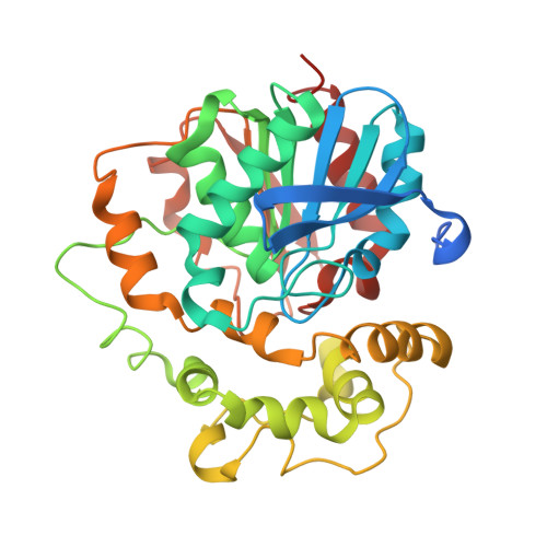
    Structure-based virtual screening was applied to design combinatorial libraries to discover novel and potent soluble epoxide hydrolase (sEH) inhibitors. X-ray crystal structures revealed unique interactions for a benzoxazole template in addition to the conserved hydrogen bonds with the catalytic machinery of sEH. By exploitation of the favorable binding elements, two iterations of library design based on amide coupling were employed, guided principally by the docking results of the enumerated virtual products. Biological screening of the libraries demonstrated as high as 90% hit rate, of which over two dozen compounds were single digit nanomolar sEH inhibitors by IC(50) determination. In total the library design and synthesis produced more than 300 submicromolar sEH inhibitors. In cellular systems consistent activities were demonstrated with biochemical measurements. The SAR understanding of the benzoxazole template provides valuable insights into discovery of novel sEH inhibitors as therapeutic agents.


  • Organizational Affiliation
    • Pfizer Global Research and Development, 700 Chesterfield Parkway West, Chesterfield, Missouri 63017, United States. li.xing@pfizer.com

Macromolecule Content 

  • Total Structure Weight: 79.18 kDa 
  • Atom Count: 5,425 
  • Modeled Residue Count: 646 
  • Deposited Residue Count: 688 
  • Unique protein chains: 1

Macromolecules

Find similar proteins by:|  3D Structure
Entity ID: 1
MoleculeChains  Sequence LengthOrganismDetailsImage
Epoxide hydrolase 2
A, B
344Homo sapiensMutation(s): 0 
Gene Names: EPHX2
EC: 3.3.2.10 (PDB Primary Data), 3.1.3.76 (UniProt)
UniProt & NIH Common Fund Data Resources
Find proteins for P34913 (Homo sapiens)
Explore P34913 
Go to UniProtKB:  P34913
PHAROS:  P34913
GTEx:  ENSG00000120915 
Entity Groups
Sequence Clusters30% Identity50% Identity70% Identity90% Identity95% Identity100% Identity
UniProt GroupP34913
Sequence Annotations
Expand
Reference Sequence

Small Molecules

Experimental Data & Validation

Experimental Data

  • Method: X-RAY DIFFRACTION
  • Resolution: 2.60 Å
  • R-Value Free:  0.333 (Depositor), 0.324 (DCC) 
  • R-Value Work:  0.279 (Depositor), 0.273 (DCC) 
  • R-Value Observed: 0.279 (Depositor) 
Space Group: P 1 21 1
Unit Cell:
Length ( Å )Angle ( ˚ )
a = 46.523α = 90
b = 79.902β = 90.223
c = 89.318γ = 90
Software Package:
Software NamePurpose
DENZOdata reduction
SCALEPACKdata scaling
CNSrefinement
PDB_EXTRACTdata extraction
MAR345dtbdata collection
X-PLORphasing
X-PLORrefinement

Structure Validation

View Full Validation Report


Ligand Structure Quality Assessment 


Entry History 

Deposition Data

Revision History  (Full details and data files)

  • Version 1.0: 2011-04-06
    Type: Initial release
  • Version 1.1: 2011-07-13
    Changes: Version format compliance
  • Version 1.2: 2021-02-24
    Changes: Database references, Derived calculations
  • Version 1.3: 2023-09-06
    Changes: Data collection, Database references, Refinement description