


Crystal structure of an atypical two-cysteine peroxiredoxin (SAOUHSC_01822) from Staphylococcus aureus NCTC8325
Bhattacharyya, S., Dutta, D., Ghosh, A.K., Das, A.K.To be published.
Experimental Data Snapshot
Starting Model: experimental
View more details
Entity ID: 1 | |||||
|---|---|---|---|---|---|
| Molecule | Chains | Sequence Length | Organism | Details | Image |
| Probable thiol peroxidase | 166 | Staphylococcus aureus subsp. aureus NCTC 8325 | Mutation(s): 0 Gene Names: SAOUHSC_01822, tpx EC: 1.11.1 (PDB Primary Data), 1.11.1.24 (UniProt) | 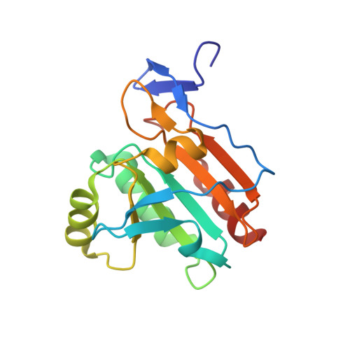 | |
UniProt | |||||
Entity Groups | |||||
| Sequence Clusters | 30% Identity50% Identity70% Identity90% Identity95% Identity100% Identity | ||||
| UniProt Group | Q2FXL3 | ||||
Sequence AnnotationsExpand | |||||
Reference Sequence | |||||
| Ligands 4 Unique | |||||
|---|---|---|---|---|---|
| ID | Chains | Name / Formula / InChI Key | 2D Diagram | 3D Interactions | |
| PG4 Download:Ideal Coordinates CCD File | F [auth A] G [auth A] H [auth A] I [auth A] K [auth B] | TETRAETHYLENE GLYCOL C8 H18 O5 UWHCKJMYHZGTIT-UHFFFAOYSA-N |  | ||
| DTU Download:Ideal Coordinates CCD File | W [auth D] | (2R,3S)-1,4-DIMERCAPTOBUTANE-2,3-DIOL C4 H10 O2 S2 VHJLVAABSRFDPM-ZXZARUISSA-N |  | ||
| DTV Download:Ideal Coordinates CCD File | J [auth A] | (2S,3S)-1,4-DIMERCAPTOBUTANE-2,3-DIOL C4 H10 O2 S2 VHJLVAABSRFDPM-QWWZWVQMSA-N |  | ||
| SO4 Download:Ideal Coordinates CCD File | E [auth A], N [auth C] | SULFATE ION O4 S QAOWNCQODCNURD-UHFFFAOYSA-L |  | ||
| Length ( Å ) | Angle ( ˚ ) |
|---|---|
| a = 43.5 | α = 90 |
| b = 149.356 | β = 104.43 |
| c = 73.736 | γ = 90 |
| Software Name | Purpose |
|---|---|
| SCALA | data scaling |
| REFMAC | refinement |
| PDB_EXTRACT | data extraction |
| StructureStudio | data collection |
| XDS | data reduction |
| MOLREP | phasing |


