3P16 | pdb_00003p16

Crystal structure of DNA polymerase III sliding clamp


Experimental Data Snapshot

  • Method: X-RAY DIFFRACTION
  • Resolution: 2.89 Å
  • R-Value Free: 
    0.287 (Depositor), 0.259 (DCC) 
  • R-Value Work: 
    0.260 (Depositor) 
  • R-Value Observed: 
    0.261 (Depositor) 

Starting Model: experimental
View more details

wwPDB Validation 3D Report Full Report

Validation slider image for 3P16

This is version 1.1 of the entry. See complete history

Literature

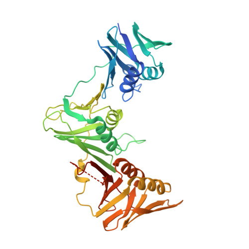
Crystal structure of DNA polymerase III beta sliding clamp from Mycobacterium tuberculosis.

Gui, W.J.Lin, S.Q.Chen, Y.Y.Zhang, X.E.Bi, L.J.Jiang, T.

(2011) Biochem Biophys Res Commun 405: 272-277

  • DOI: https://doi.org/10.1016/j.bbrc.2011.01.027
  • Primary Citation Related Structures: 
    3P16

  • PubMed Abstract: 

    The sliding clamp is a key component of DNA polymerase III (Pol III) required for genome replication. It is known to function with diverse DNA repair proteins and cell cycle-control proteins, making it a potential drug target. To extend our understanding of the structure/function relationship of the sliding clamp, we solved the crystal structure of the sliding clamp from Mycobacterium tuberculosis (M. tuberculosis), a human pathogen that causes most cases of tuberculosis (TB). The sliding clamp from M. tuberculosis forms a ring-shaped head-to-tail dimer with three domains per subunit. Each domain contains two α helices in the inner ring that lie against two β sheets in the outer ring. Previous studies have indicated that many Escherichia coli clamp-binding proteins have a conserved LF sequence, which is critical for binding to the hydrophobic region of the sliding clamp. Here, we analyzed the binding affinities of the M. tuberculosis sliding clamp and peptides derived from the α and δ subunits of Pol III, which indicated that the LF motif also plays an important role in the binding of the α and δ subunits to the sliding clamp of M. tuberculosis.


  • Organizational Affiliation
    • National Laboratory of Biomacromolecules, Institute of Biophysics, Chinese Academy of Sciences, 15 Datun Road, Chaoyang District, Beijing 100101, China.

Macromolecule Content 

  • Total Structure Weight: 257.88 kDa 
  • Atom Count: 16,587 
  • Modeled Residue Count: 2,238 
  • Deposited Residue Count: 2,448 
  • Unique protein chains: 1

Macromolecules

Find similar proteins by:|  3D Structure
Entity ID: 1
MoleculeChains  Sequence LengthOrganismDetailsImage
DNA polymerase III subunit beta
A, B, C, D, E
A, B, C, D, E, F
408Mycobacterium tuberculosis H37RvMutation(s): 0 
Gene Names: dnaNRv0002MT0002MTV029.02MTCY10H4.0
EC: 2.7.7.7
UniProt
Find proteins for P9WNU1 (Mycobacterium tuberculosis (strain ATCC 25618 / H37Rv))
Explore P9WNU1 
Go to UniProtKB:  P9WNU1
Entity Groups
Sequence Clusters30% Identity50% Identity70% Identity90% Identity95% Identity100% Identity
UniProt GroupP9WNU1
Sequence Annotations
Expand
Reference Sequence

Experimental Data & Validation

Experimental Data

  • Method: X-RAY DIFFRACTION
  • Resolution: 2.89 Å
  • R-Value Free:  0.287 (Depositor), 0.259 (DCC) 
  • R-Value Work:  0.260 (Depositor) 
  • R-Value Observed: 0.261 (Depositor) 
Space Group: P 1 21 1
Unit Cell:
Length ( Å )Angle ( ˚ )
a = 88.05α = 90
b = 132.974β = 109.05
c = 113.777γ = 90
Software Package:
Software NamePurpose
HKL-2000data collection
PHASERphasing
REFMACrefinement
HKL-2000data reduction
HKL-2000data scaling

Structure Validation

View Full Validation Report


Entry History 

Deposition Data

Revision History  (Full details and data files)

  • Version 1.0: 2011-08-24
    Type: Initial release
  • Version 1.1: 2023-11-01
    Changes: Data collection, Database references, Refinement description