3P03 | pdb_00003p03

Crystal structure of BetP-G153D with choline bound


Experimental Data Snapshot

  • Method: X-RAY DIFFRACTION
  • Resolution: 3.35 Å
  • R-Value Free: 
    0.300 (Depositor), 0.316 (DCC) 
  • R-Value Work: 
    0.245 (Depositor), 0.278 (DCC) 
  • R-Value Observed: 
    0.250 (Depositor) 

wwPDB Validation 3D Report Full Report

Validation slider image for 3P03

This is version 1.2 of the entry. See complete history

Literature

Substrate specificity and ion coupling in the Na(+)/betaine symporter BetP.

Perez, C.Koshy, C.Ressl, S.Nicklisch, S.Kramer, R.Ziegler, C.

(2011) EMBO J 30: 1221-1229

  • DOI: https://doi.org/10.1038/emboj.2011.46
  • Primary Citation Related Structures: 
    3P03

  • PubMed Abstract: 

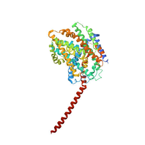
    BetP is an Na(+)-coupled betaine-specific transporter of the betaine-choline-carnitine (BCC) transporter family involved in the response to hyperosmotic stress. The crystal structure of BetP revealed an overall fold of two inverted structurally related repeats (LeuT-fold) that BetP shares with other sequence-unrelated Na(+)-coupled symporters. Numerous structures of LeuT-fold transporters in distinct conformational states have contributed substantially to our understanding of the alternating access mechanism of transport. Nevertheless, coupling of substrate and co-transported ion fluxes has not been structurally corroborated to the same extent. We converted BetP by a single-point mutation--glycine to aspartate--into an H(+)-coupled choline-specific transporter and solved the crystal structure of this mutant in complex with choline. The structure of BetP-G153D demonstrates a new inward-facing open conformation for BetP. Choline binding to a location close to the second, low-affinity sodium-binding site (Na2) of LeuT-fold transporters is facilitated by the introduced aspartate. Our data confirm the importance of a cation-binding site in BetP, playing a key role in a proposed molecular mechanism of Na(+) and H(+) coupling in BCC transporters.


  • Organizational Affiliation
    • Department of Structural Biology, Max-Planck Institute of Biophysics, Frankfurt am Main, Germany.

Macromolecule Content 

  • Total Structure Weight: 183.29 kDa 
  • Atom Count: 11,349 
  • Modeled Residue Count: 1,488 
  • Deposited Residue Count: 1,698 
  • Unique protein chains: 1

Macromolecules

Find similar proteins by:|  3D Structure
Entity ID: 1
MoleculeChains  Sequence LengthOrganismDetailsImage
Glycine betaine transporter BetP
A, B, C
566Corynebacterium glutamicumMutation(s): 4 
Gene Names: betPCgl0892cg1016
Membrane Entity: Yes 
UniProt
Find proteins for P54582 (Corynebacterium glutamicum (strain ATCC 13032 / DSM 20300 / JCM 1318 / BCRC 11384 / CCUG 27702 / LMG 3730 / NBRC 12168 / NCIMB 10025 / NRRL B-2784 / 534))
Explore P54582 
Go to UniProtKB:  P54582
Entity Groups
Sequence Clusters30% Identity50% Identity70% Identity90% Identity95% Identity100% Identity
UniProt GroupP54582
Sequence Annotations
Expand
Reference Sequence

Small Molecules

Ligands 1 Unique
IDChains Name / Formula / InChI Key2D Diagram3D Interactions
CHT

Query on CHT


Download:Ideal Coordinates CCD File
D [auth C]CHOLINE ION
C5 H14 N O
OEYIOHPDSNJKLS-UHFFFAOYSA-N

Experimental Data & Validation

Experimental Data

  • Method: X-RAY DIFFRACTION
  • Resolution: 3.35 Å
  • R-Value Free:  0.300 (Depositor), 0.316 (DCC) 
  • R-Value Work:  0.245 (Depositor), 0.278 (DCC) 
  • R-Value Observed: 0.250 (Depositor) 
Space Group: P 21 21 21
Unit Cell:
Length ( Å )Angle ( ˚ )
a = 117.56α = 90
b = 129.31β = 90
c = 183.14γ = 90
Software Package:
Software NamePurpose
XSCALEdata scaling
PHASERphasing
PHENIXrefinement
PDB_EXTRACTdata extraction
XDSdata scaling
XDSdata reduction

Structure Validation

View Full Validation Report


Entry History 

Deposition Data

Revision History  (Full details and data files)

  • Version 1.0: 2011-03-30
    Type: Initial release
  • Version 1.1: 2011-07-13
    Changes: Version format compliance
  • Version 1.2: 2024-02-21
    Changes: Data collection, Database references, Derived calculations