3OZD

Crystal Structure of human 5'-deoxy-5'-methyladenosine phosphorylase in complex with pCl-phenylthioDADMeImmA


Experimental Data Snapshot

  • Method: X-RAY DIFFRACTION
  • Resolution: 2.10 Å
  • R-Value Free: 
    0.257 (Depositor), 0.260 (DCC) 
  • R-Value Work: 
    0.200 (Depositor), 0.210 (DCC) 
  • R-Value Observed: 
    0.202 (Depositor) 

wwPDB Validation   3D Report Full Report


Ligand Structure Quality Assessment 

Created with Raphaël 2.3.0Worse 01 BetterLigand structure goodness of fit to experimental dataBest fitted 4CTClick on this verticalbar to view details

This is version 1.2 of the entry. See complete history


Literature

Crystal Structure of human 5'-deoxy-5'-methyladenosine phosphorylase

Ho, M.Guan, R.Almo, S.C.Schramm, V.L.

To be published.

Macromolecules
Find similar proteins by:  (by identity cutoff)  |  3D Structure
Entity ID: 1
MoleculeChains Sequence LengthOrganismDetailsImage
S-methyl-5'-thioadenosine phosphorylase
A, B
283Homo sapiensMutation(s): 0 
Gene Names: MTAPMSAP
EC: 2.4.2.28
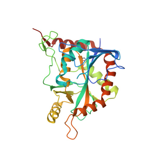
UniProt & NIH Common Fund Data Resources
Find proteins for Q13126 (Homo sapiens)
Explore Q13126 
Go to UniProtKB:  Q13126
PHAROS:  Q13126
GTEx:  ENSG00000099810 
Entity Groups  
Sequence Clusters30% Identity50% Identity70% Identity90% Identity95% Identity100% Identity
UniProt GroupQ13126
Sequence Annotations
Expand
  • Reference Sequence
Small Molecules
Ligands 1 Unique
IDChains Name / Formula / InChI Key2D Diagram3D Interactions
4CT
Query on 4CT

Download Ideal Coordinates CCD File 
C [auth A],
D [auth B]
(3R,4S)-1-[(4-amino-5H-pyrrolo[3,2-d]pyrimidin-7-yl)methyl]-4-{[(4-chlorophenyl)sulfanyl]methyl}pyrrolidin-3-ol
C18 H20 Cl N5 O S
MZZBHZOHYGEGEE-DOMZBBRYSA-N
Binding Affinity Annotations 
IDSourceBinding Affinity
4CT BindingDB:  3OZD Kd: 1.00e-2 (nM) from 1 assay(s)
Experimental Data & Validation

Experimental Data

  • Method: X-RAY DIFFRACTION
  • Resolution: 2.10 Å
  • R-Value Free:  0.257 (Depositor), 0.260 (DCC) 
  • R-Value Work:  0.200 (Depositor), 0.210 (DCC) 
  • R-Value Observed: 0.202 (Depositor) 
Space Group: P 63
Unit Cell:
Length ( Å )Angle ( ˚ )
a = 121.539α = 90
b = 121.539β = 90
c = 87.661γ = 120
Software Package:
Software NamePurpose
DENZOdata reduction
SCALEPACKdata scaling
MOLREPphasing
REFMACrefinement
PDB_EXTRACTdata extraction

Structure Validation

View Full Validation Report


Ligand Structure Quality Assessment 

Created with Raphaël 2.3.0Worse 01 BetterLigand structure goodness of fit to experimental dataBest fitted 4CTClick on this verticalbar to view details

Entry History 

Deposition Data

Revision History  (Full details and data files)

  • Version 1.0: 2011-09-28
    Type: Initial release
  • Version 1.1: 2017-11-08
    Changes: Refinement description
  • Version 1.2: 2024-11-27
    Changes: Data collection, Database references, Derived calculations, Structure summary