3OZ1 | pdb_00003oz1

cIAP1-BIR3 domain in complex with the Smac-mimetic compound Smac066


Experimental Data Snapshot

  • Method: X-RAY DIFFRACTION
  • Resolution: 3.00 Å
  • R-Value Free: 
    0.257 (Depositor), 0.277 (DCC) 
  • R-Value Work: 
    0.192 (Depositor), 0.210 (DCC) 
  • R-Value Observed: 
    0.195 (Depositor) 

wwPDB Validation 3D Report Full Report

Validation slider image for 3OZ1

Ligand Structure Quality Assessment 


This is version 1.2 of the entry. See complete history

Literature

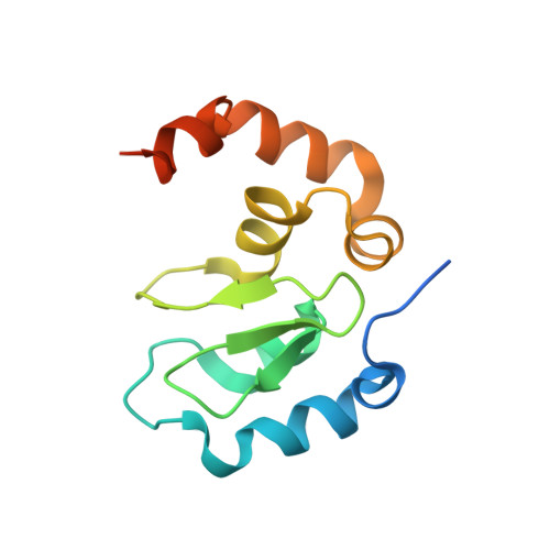
Recognition of Smac-mimetic compounds by the BIR3 domain of cIAP1

Cossu, F.Malvezzi, F.Canevari, G.Mastrangelo, E.Lecis, D.Delia, D.Seneci, P.Scolastico, C.Bolognesi, M.Milani, M.

(2010) Protein Sci 

  • DOI: https://doi.org/10.1002/pro.523
  • Primary Citation Related Structures: 
    3MUP, 3OZ1

  • PubMed Abstract: 

    Inhibitor of apoptosis proteins (IAPs) are negative regulators of apoptosis. As IAPs are overexpressed in many tumors, where they confer chemoresistance, small molecules inactivating IAPs have been proposed as anticancer agents. Accordingly, a number of IAP-binding pro-apoptotic compounds that mimic the sequence corresponding to the N-terminal tetrapeptide of Smac/DIABLO, the natural endogenous IAPs inhibitor, have been developed. Here, we report the crystal structures of the BIR3 domain of cIAP1 in complex with Smac037, a Smac-mimetic known to bind potently to the XIAP-BIR3 domain and to induce degradation of cIAP1, and in complex with the novel Smac-mimetic compound Smac066. Thermal stability and fluorescence polarization assays show the stabilizing effect and the high affinity of both Smac037 and Smac066 for cIAP1- and cIAP2-BIR3 domains.


  • Organizational Affiliation
    • Dipartimento di Scienze Biomolecolari e Biotecnologie, Università di Milano, Via Celoria 26, I-20133, Milano, Italy.

Macromolecule Content 

  • Total Structure Weight: 60.26 kDa 
  • Atom Count: 3,579 
  • Modeled Residue Count: 412 
  • Deposited Residue Count: 488 
  • Unique protein chains: 1

Macromolecules

Find similar proteins by:|  3D Structure
Entity ID: 1
MoleculeChains  Sequence LengthOrganismDetailsImage
Baculoviral IAP repeat-containing protein 2
A, B, C, D
122Homo sapiensMutation(s): 0 
Gene Names: BIRC2API1IAP2MIHBRNF48
EC: 2.3.2.27
UniProt & NIH Common Fund Data Resources
Find proteins for Q13490 (Homo sapiens)
Explore Q13490 
Go to UniProtKB:  Q13490
PHAROS:  Q13490
GTEx:  ENSG00000110330 
Entity Groups
Sequence Clusters30% Identity50% Identity70% Identity90% Identity95% Identity100% Identity
UniProt GroupQ13490
Sequence Annotations
Expand
Reference Sequence

Small Molecules

Ligands 2 Unique
IDChains Name / Formula / InChI Key2D Diagram3D Interactions
BMB

Query on BMB


Download:Ideal Coordinates CCD File
F [auth A],
H [auth B],
J [auth C],
L [auth D]
(3S,6S,7R,9aS)-7-[2-(benzylamino)ethyl]-N-(diphenylmethyl)-6-{[(2S)-2-(methylamino)butanoyl]amino}-5-oxooctahydro-1H-pyrrolo[1,2-a]azepine-3-carboxamide
C37 H47 N5 O3
NVNDSZWHCFDEAR-UCDLYESDSA-N
ZN

Query on ZN


Download:Ideal Coordinates CCD File
E [auth A],
G [auth B],
I [auth C],
K [auth D]
ZINC ION
Zn
PTFCDOFLOPIGGS-UHFFFAOYSA-N

Experimental Data & Validation

Experimental Data

  • Method: X-RAY DIFFRACTION
  • Resolution: 3.00 Å
  • R-Value Free:  0.257 (Depositor), 0.277 (DCC) 
  • R-Value Work:  0.192 (Depositor), 0.210 (DCC) 
  • R-Value Observed: 0.195 (Depositor) 
Space Group: C 2 2 2
Unit Cell:
Length ( Å )Angle ( ˚ )
a = 113.589α = 90
b = 114.63β = 90
c = 92.962γ = 90
Software Package:
Software NamePurpose
DNAdata collection
PHASERphasing
BUSTERrefinement
MOSFLMdata reduction
SCALAdata scaling

Structure Validation

View Full Validation Report


Ligand Structure Quality Assessment 


Entry History 

Deposition Data

Revision History  (Full details and data files)

  • Version 1.0: 2010-11-24
    Type: Initial release
  • Version 1.1: 2011-07-13
    Changes: Version format compliance
  • Version 1.2: 2024-03-20
    Changes: Data collection, Database references, Derived calculations