3OYZ | pdb_00003oyz

Haloferax volcanii Malate Synthase Pyruvate/Acetyl-CoA Ternary Complex


Experimental Data Snapshot

  • Method: X-RAY DIFFRACTION
  • Resolution: 1.95 Å
  • R-Value Free: 
    0.239 (Depositor), 0.239 (DCC) 
  • R-Value Work: 
    0.203 (Depositor), 0.204 (DCC) 
  • R-Value Observed: 
    0.205 (Depositor) 

Starting Model: experimental
View more details

wwPDB Validation 3D Report Full Report

Validation slider image for 3OYZ

Ligand Structure Quality Assessment 


This is version 2.1 of the entry. See complete history

Literature

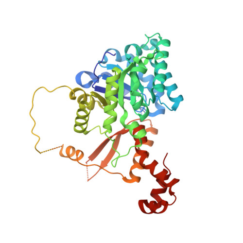
Crystal structures of a halophilic archaeal malate synthase from Haloferax volcanii and comparisons with isoforms A and G.

Bracken, C.D.Neighbor, A.M.Lamlenn, K.K.Thomas, G.C.Schubert, H.L.Whitby, F.G.Howard, B.R.

(2011) BMC Struct Biol 11: 23-23

  • DOI: https://doi.org/10.1186/1472-6807-11-23
  • Primary Citation Related Structures: 
    3OYX, 3OYZ, 3PUG

  • PubMed Abstract: 

    Malate synthase, one of the two enzymes unique to the glyoxylate cycle, is found in all three domains of life, and is crucial to the utilization of two-carbon compounds for net biosynthetic pathways such as gluconeogenesis. In addition to the main isoforms A and G, so named because of their differential expression in E. coli grown on either acetate or glycolate respectively, a third distinct isoform has been identified. These three isoforms differ considerably in size and sequence conservation. The A isoform (MSA) comprises ~530 residues, the G isoform (MSG) is ~730 residues, and this third isoform (MSH-halophilic) is ~430 residues in length. Both isoforms A and G have been structurally characterized in detail, but no structures have been reported for the H isoform which has been found thus far only in members of the halophilic Archaea. We have solved the structure of a malate synthase H (MSH) isoform member from Haloferax volcanii in complex with glyoxylate at 2.51 Å resolution, and also as a ternary complex with acetyl-coenzyme A and pyruvate at 1.95 Å. Like the A and G isoforms, MSH is based on a β8/α8 (TIM) barrel. Unlike previously solved malate synthase structures which are all monomeric, this enzyme is found in the native state as a trimer/hexamer equilibrium. Compared to isoforms A and G, MSH displays deletion of an N-terminal domain and a smaller deletion at the C-terminus. The MSH active site is closely superimposable with those of MSA and MSG, with the ternary complex indicating a nucleophilic attack on pyruvate by the enolate intermediate of acetyl-coenzyme A. The reported structures of MSH from Haloferax volcanii allow a detailed analysis and comparison with previously solved structures of isoforms A and G. These structural comparisons provide insight into evolutionary relationships among these isoforms, and also indicate that despite the size and sequence variation, and the truncated C-terminal domain of the H isoform, the catalytic mechanism is conserved. Sequence analysis in light of the structure indicates that additional members of isoform H likely exist in the databases but have been misannotated.


  • Organizational Affiliation
    • Department of Physical Science, Southern Utah University, Cedar City, UT 84720-2470, USA.

Macromolecule Content 

  • Total Structure Weight: 49.34 kDa 
  • Atom Count: 3,197 
  • Modeled Residue Count: 376 
  • Deposited Residue Count: 433 
  • Unique protein chains: 1

Macromolecules

Find similar proteins by:|  3D Structure
Entity ID: 1
MoleculeChains  Sequence LengthOrganismDetailsImage
Malate Synthase433Haloferax volcanii DS2Mutation(s): 0 
EC: 2.3.3.9
UniProt
Find proteins for D4GTL2 (Haloferax volcanii (strain ATCC 29605 / DSM 3757 / JCM 8879 / NBRC 14742 / NCIMB 2012 / VKM B-1768 / DS2))
Explore D4GTL2 
Go to UniProtKB:  D4GTL2
Entity Groups
Sequence Clusters30% Identity50% Identity70% Identity90% Identity95% Identity100% Identity
UniProt GroupD4GTL2
Sequence Annotations
Expand
Reference Sequence

Small Molecules

Ligands 6 Unique
IDChains Name / Formula / InChI Key2D Diagram3D Interactions
ACO

Query on ACO


Download:Ideal Coordinates CCD File
B [auth A]ACETYL COENZYME *A
C23 H38 N7 O17 P3 S
ZSLZBFCDCINBPY-ZSJPKINUSA-N
PO4

Query on PO4


Download:Ideal Coordinates CCD File
L [auth A]PHOSPHATE ION
O4 P
NBIIXXVUZAFLBC-UHFFFAOYSA-K
PYR

Query on PYR


Download:Ideal Coordinates CCD File
C [auth A]PYRUVIC ACID
C3 H4 O3
LCTONWCANYUPML-UHFFFAOYSA-N
K

Query on K


Download:Ideal Coordinates CCD File
E [auth A],
F [auth A],
G [auth A]
POTASSIUM ION
K
NPYPAHLBTDXSSS-UHFFFAOYSA-N
CL

Query on CL


Download:Ideal Coordinates CCD File
H [auth A],
I [auth A],
J [auth A],
K [auth A]
CHLORIDE ION
Cl
VEXZGXHMUGYJMC-UHFFFAOYSA-M
MG

Query on MG


Download:Ideal Coordinates CCD File
D [auth A]MAGNESIUM ION
Mg
JLVVSXFLKOJNIY-UHFFFAOYSA-N

Experimental Data & Validation

Experimental Data

  • Method: X-RAY DIFFRACTION
  • Resolution: 1.95 Å
  • R-Value Free:  0.239 (Depositor), 0.239 (DCC) 
  • R-Value Work:  0.203 (Depositor), 0.204 (DCC) 
  • R-Value Observed: 0.205 (Depositor) 
Space Group: H 3 2
Unit Cell:
Length ( Å )Angle ( ˚ )
a = 154.788α = 90
b = 154.788β = 90
c = 142.144γ = 120
Software Package:
Software NamePurpose
HKL-2000data collection
SOLVEphasing
REFMACrefinement
HKL-2000data reduction
HKL-2000data scaling

Structure Validation

View Full Validation Report


Ligand Structure Quality Assessment 


Entry History 

Deposition Data

Revision History  (Full details and data files)

  • Version 1.0: 2011-06-01
    Type: Initial release
  • Version 1.1: 2011-07-13
    Changes: Version format compliance
  • Version 1.2: 2018-01-24
    Changes: Structure summary
  • Version 2.0: 2023-11-15
    Changes: Atomic model, Data collection, Database references, Derived calculations
  • Version 2.1: 2024-04-03
    Changes: Refinement description