3OYO | pdb_00003oyo

Crystal structure of hemopexin fold protein CP4 from cow pea


Experimental Data Snapshot

  • Method: X-RAY DIFFRACTION
  • Resolution: 2.10 Å
  • R-Value Free: 
    0.246 (Depositor), 0.243 (DCC) 
  • R-Value Work: 
    0.205 (Depositor), 0.202 (DCC) 
  • R-Value Observed: 
    0.209 (Depositor) 

Starting Model: experimental
View more details

wwPDB Validation 3D Report Full Report

Validation slider image for 3OYO

This is version 1.3 of the entry. See complete history

Literature

The structure of a haemopexin-fold protein from cow pea (Vigna unguiculata) suggests functional diversity of haemopexins in plants

Gaur, V.Chanana, V.Jain, A.Salunke, D.M.

(2011) Acta Crystallogr Sect F Struct Biol Cryst Commun 67: 193-200

  • DOI: https://doi.org/10.1107/S1744309110051250
  • Primary Citation Related Structures: 
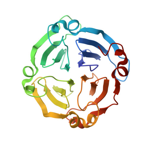
    3OYO

  • PubMed Abstract: 

    The haemopexin fold is present in almost all life forms and is utilized for carrying out diverse physiological functions. The structure of CP4, a haemopexin-fold protein from cow pea (Vigna unguiculata), was determined at 2.1 Å resolution. The protein exists as a monomer both in solution and in the crystal. The structure revealed a typical four-bladed β-propeller topology. The protein exhibits 42% sequence similarity to LS-24 from Lathyrus sativus, with substantial differences in the surface-charge distribution and in the oligomeric state. A structure-based sequence analysis of haemopexin-fold proteins of plant and mammalian origin established a sequence signature associated with the haemopexin motif. This signature sequence enabled the identification of other proteins with possible haemopexin-like topology of both plant and animal origin. Although CP4 shares a structural fold with LS-24 and other haemopexins, biochemical studies indicated possible functional differences between CP4 and LS-24. While both of these proteins exhibit spermine-binding potential, CP4 does not bind to haem, unlike LS-24.


  • Organizational Affiliation
    • National Institute of Immunology, Aruna Asaf Ali Marg, New Delhi, India.

Macromolecule Content 

  • Total Structure Weight: 49.68 kDa 
  • Atom Count: 3,692 
  • Modeled Residue Count: 450 
  • Deposited Residue Count: 450 
  • Unique protein chains: 1

Macromolecules

Find similar proteins by:|  3D Structure
Entity ID: 1
MoleculeChains  Sequence LengthOrganismDetailsImage
hemopexin fold protein CP4
A, B
225Vigna unguiculataMutation(s): 0 
UniProt
Find proteins for F2Z290 (Vigna unguiculata)
Explore F2Z290 
Go to UniProtKB:  F2Z290
Entity Groups
Sequence Clusters30% Identity50% Identity70% Identity90% Identity95% Identity100% Identity
UniProt GroupF2Z290
Sequence Annotations
Expand
Reference Sequence

Experimental Data & Validation

Experimental Data

  • Method: X-RAY DIFFRACTION
  • Resolution: 2.10 Å
  • R-Value Free:  0.246 (Depositor), 0.243 (DCC) 
  • R-Value Work:  0.205 (Depositor), 0.202 (DCC) 
  • R-Value Observed: 0.209 (Depositor) 
Space Group: C 1 2 1
Unit Cell:
Length ( Å )Angle ( ˚ )
a = 125.087α = 90
b = 60.138β = 111.18
c = 67.574γ = 90
Software Package:
Software NamePurpose
MAR345dtbdata collection
AMoREphasing
REFMACrefinement
AUTOMARdata reduction

Structure Validation

View Full Validation Report


Entry History 

Deposition Data

Revision History  (Full details and data files)

  • Version 1.0: 2011-02-09
    Type: Initial release
  • Version 1.1: 2011-07-13
    Changes: Version format compliance
  • Version 1.2: 2014-03-26
    Changes: Database references
  • Version 1.3: 2023-11-01
    Changes: Data collection, Database references, Derived calculations, Refinement description