3OTJ | pdb_00003otj

A Crystal Structure of Trypsin Complexed with BPTI (Bovine Pancreatic Trypsin Inhibitor) by X-ray/Neutron Joint Refinement


Experimental Data Snapshot

  • Method: NEUTRON DIFFRACTION
  • Resolution: 2.15 Å
  • R-Value Free: 
    0.226 (Depositor) 
  • R-Value Work: 
    0.209 (Depositor) 

  • Method: X-RAY DIFFRACTION
  • Resolution: 1.60 Å
  • R-Value Free: 
    0.209 (Depositor), 0.197 (DCC) 
  • R-Value Work: 
    0.198 (Depositor), 0.182 (DCC) 

wwPDB Validation 3D Report Full Report

Validation slider image for 3OTJ

This is version 1.5 of the entry. See complete history

Literature

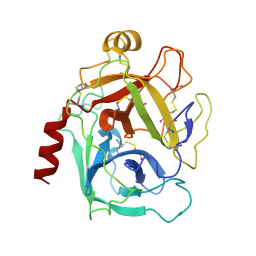
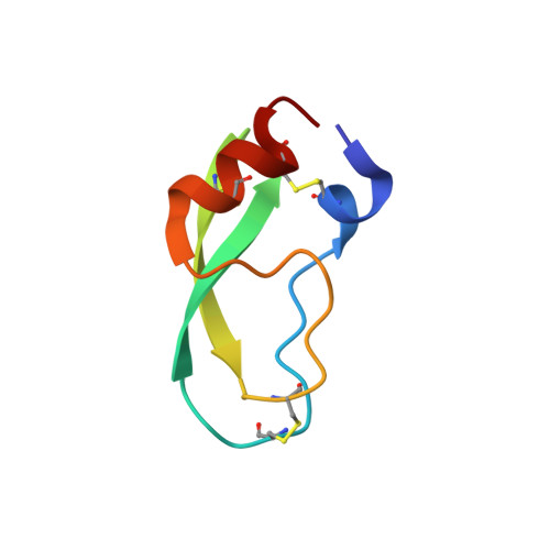
X-ray and neutron protein crystallographic analysis of the trypsin-BPTI complex.

Kawamura, K.Yamada, T.Kurihara, K.Tamada, T.Kuroki, R.Tanaka, I.Takahashi, H.Niimura, N.

(2011) Acta Crystallogr D Biol Crystallogr 67: 140-148

  • DOI: https://doi.org/10.1107/S0907444910053382
  • Primary Citation Related Structures: 
    3OTJ

  • PubMed Abstract: 

    In this work, the crystal structure of the β-trypsin-bovine pancreatic trypsin inhibitor (BPTI) complex was refined and the D and H atoms in the complex were identified using data from both 1.6 Å resolution X-ray diffraction and 2.15 Å resolution neutron diffraction. After crystallization in an H(2)O solution, the sample crystal was soaked in a D(2)O solution for about two weeks. The protonation states of the catalytic triad (Asp102, His57 and Ser195) were observed. These results confirmed that the nucleophilic reactivity of the hydroxyl group of Ser195 was increased by forming a hydrogen bond with His57. According to structural analysis, the trypsin-BPTI interfaces located at the scissile peptide and the active sites were inaccessible to solvent water, and the amide H atoms of P2' Arg17/I, Gly216/E and Gly193/E at the binding interface were protected from H/D exchange. In contrast, both the amide H atom of P1' Ala16/I of the scissile peptide bond P1-P1' and the H atom between His57 N(ℇ2) and Ser195 O(γ) were replaced by D atoms. The hydrogen-bond networks at the S1 pocket were also confirmed and discussed from the viewpoint of substrate recognition. Moreover, the first neutron crystallographic structure of the Michaelis complex state of trypsin-BPTI is presented.


  • Organizational Affiliation
    • Institute of Applied Beam Science, Graduate School of Science and Engineering, Ibaraki University, 4-12-1 Naka-Narusawa, Hitachi, Ibaraki 316-8511, Japan.

Macromolecule Content 

  • Total Structure Weight: 30.37 kDa 
  • Atom Count: 2,229 
  • Modeled Residue Count: 281 
  • Deposited Residue Count: 281 
  • Unique protein chains: 2

Macromolecules

Find similar proteins by:|  3D Structure
Entity ID: 1
MoleculeChains  Sequence LengthOrganismDetailsImage
Cationic trypsinA [auth E]223Bos taurusMutation(s): 0 
EC: 3.4.21.4
UniProt
Find proteins for P00760 (Bos taurus)
Explore P00760 
Go to UniProtKB:  P00760
Entity Groups
Sequence Clusters30% Identity50% Identity70% Identity90% Identity95% Identity100% Identity
UniProt GroupP00760
Sequence Annotations
Expand
Reference Sequence
Find similar proteins by:|  3D Structure
Entity ID: 2
MoleculeChains  Sequence LengthOrganismDetailsImage
Pancreatic trypsin inhibitorB [auth I]58Bos taurusMutation(s): 0 
UniProt
Find proteins for P00974 (Bos taurus)
Explore P00974 
Go to UniProtKB:  P00974
Entity Groups
Sequence Clusters30% Identity50% Identity70% Identity90% Identity95% Identity100% Identity
UniProt GroupP00974
Sequence Annotations
Expand
Reference Sequence

Experimental Data & Validation

Experimental Data

  • Method: NEUTRON DIFFRACTION
  • Resolution: 2.15 Å
  • R-Value Free:  0.226 (Depositor) 
  • R-Value Work:  0.209 (Depositor) 
  • Method: X-RAY DIFFRACTION
  • Resolution: 1.60 Å
  • R-Value Free:  0.209 (Depositor), 0.197 (DCC) 
  • R-Value Work:  0.198 (Depositor), 0.182 (DCC) 
Space Group: I 2 2 2
Unit Cell:
Length ( Å )Angle ( ˚ )
a = 75.604α = 90
b = 85.361β = 90
c = 122.551γ = 90
Software Package:
Software NamePurpose
nCNSrefinement
DENZOdata reduction
SCALEPACKdata scaling
nCNSphasing

Structure Validation

View Full Validation Report


Entry History 

Deposition Data

Revision History  (Full details and data files)

  • Version 1.0: 2011-01-26
    Type: Initial release
  • Version 1.1: 2011-07-13
    Changes: Version format compliance
  • Version 1.2: 2011-07-27
    Changes: Data collection
  • Version 1.3: 2011-08-21
    Changes: Other
  • Version 1.4: 2017-11-08
    Changes: Refinement description
  • Version 1.5: 2024-10-16
    Changes: Data collection, Database references, Derived calculations, Structure summary