


Crystal Structure of The Complex of Group 1 Phospholipase A2 With Atropin At 1.5 A Resolution
Shukla, P.K., Kaushik, S., Sinha, M., Bhushan, A., Kaur, P., Sharma, S., Singh, T.P.To be published.
Experimental Data Snapshot
Starting Model: experimental
View more details
Entity ID: 1 | |||||
|---|---|---|---|---|---|
| Molecule | Chains | Sequence Length | Organism | Details | Image |
| Phospholipase A2 isoform 3 | 119 | Naja sagittifera | Mutation(s): 0 EC: 3.1.1.4 | 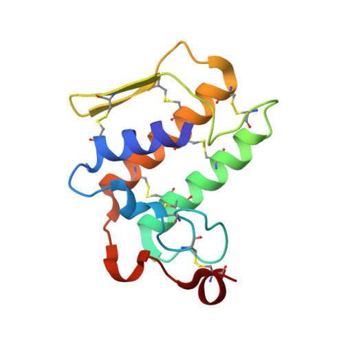 | |
UniProt | |||||
Entity Groups | |||||
| Sequence Clusters | 30% Identity50% Identity70% Identity90% Identity95% Identity100% Identity | ||||
| UniProt Group | P60045 | ||||
Sequence AnnotationsExpand | |||||
Reference Sequence | |||||
| Ligands 2 Unique | |||||
|---|---|---|---|---|---|
| ID | Chains | Name / Formula / InChI Key | 2D Diagram | 3D Interactions | |
| OIN Download:Ideal Coordinates CCD File | C [auth A] | (1R,5S)-8-METHYL-8-AZABICYCLO[3.2.1]OCT-3-YL (2R)-3-HYDROXY-2-PHENYLPROPANOATE C17 H23 N O3 RKUNBYITZUJHSG-QKPAOTATSA-N |  | ||
| CA Download:Ideal Coordinates CCD File | B [auth A] | CALCIUM ION Ca BHPQYMZQTOCNFJ-UHFFFAOYSA-N |  | ||
| Length ( Å ) | Angle ( ˚ ) |
|---|---|
| a = 42.377 | α = 90 |
| b = 42.377 | β = 90 |
| c = 65.17 | γ = 90 |
| Software Name | Purpose |
|---|---|
| HKL-2000 | data collection |
| AMoRE | phasing |
| REFMAC | refinement |
| DENZO | data reduction |
| SCALEPACK | data scaling |


