3ORF | pdb_00003orf

Crystal Structure of Dihydropteridine Reductase from Dictyostelium discoideum


Experimental Data Snapshot

  • Method: X-RAY DIFFRACTION
  • Resolution: 2.16 Å
  • R-Value Free: 
    0.240 (Depositor), 0.233 (DCC) 
  • R-Value Work: 
    0.185 (Depositor), 0.181 (DCC) 
  • R-Value Observed: 
    0.188 (Depositor) 

Starting Model: experimental
View more details

wwPDB Validation 3D Report Full Report

Validation slider image for 3ORF

Ligand Structure Quality Assessment 


This is version 1.2 of the entry. See complete history

Literature

Structural insights into the dual substrate specificities of mammalian and Dictyostelium dihydropteridine reductases toward two stereoisomers of quinonoid dihydrobiopterin

Chen, C.Kim, H.L.Zhuang, N.Seo, K.H.Park, K.H.Han, C.-D.Park, Y.S.Lee, K.H.

(2011) FEBS Lett 585: 2640-2646

  • DOI: https://doi.org/10.1016/j.febslet.2011.07.018
  • Primary Citation Related Structures: 
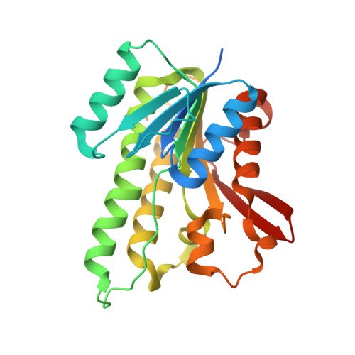
    3ORF

  • PubMed Abstract: 

    Up to now, d-threo-tetrahydrobiopterin (DH(4), dictyopterin) was detected only in Dictyostelium discoideum, while the isomer L-erythro-tetrahydrobioterin (BH(4)) is common in mammals. To elucidate the mechanism of DH(4) regeneration by D. discoideum dihydropteridine reductase (DicDHPR), we have determined the crystal structure of DicDHPR complexed with NAD(+) at 2.16 Å resolution. Significant structural differences from mammalian DHPRs are found around the coenzyme binding site, resulting in a higher K(m) value for NADH (K(m)=46.51±0.4 μM) than mammals. In addition, we have found that rat DHPR as well as DicDHPR could bind to both substrates quinonoid-BH(2) and quinonoid-DH(2) by docking calculations and have confirmed their catalytic activity by in vitro assay.


  • Organizational Affiliation
    • Division of Applied Life Science (BK21 Program), Gyeongsang National University, Jinju, Republic of Korea.

Macromolecule Content 

  • Total Structure Weight: 110.05 kDa 
  • Atom Count: 7,229 
  • Modeled Residue Count: 920 
  • Deposited Residue Count: 1,004 
  • Unique protein chains: 1

Macromolecules

Find similar proteins by:|  3D Structure
Entity ID: 1
MoleculeChains  Sequence LengthOrganismDetailsImage
Dihydropteridine reductase
A, B, C, D
251Dictyostelium discoideumMutation(s): 0 
Gene Names: DDB_G0272684qdpr
EC: 1.5.1.34
UniProt
Find proteins for Q86A17 (Dictyostelium discoideum)
Explore Q86A17 
Go to UniProtKB:  Q86A17
Entity Groups
Sequence Clusters30% Identity50% Identity70% Identity90% Identity95% Identity100% Identity
UniProt GroupQ86A17
Sequence Annotations
Expand
Reference Sequence

Experimental Data & Validation

Experimental Data

  • Method: X-RAY DIFFRACTION
  • Resolution: 2.16 Å
  • R-Value Free:  0.240 (Depositor), 0.233 (DCC) 
  • R-Value Work:  0.185 (Depositor), 0.181 (DCC) 
  • R-Value Observed: 0.188 (Depositor) 
Space Group: P 1 21 1
Unit Cell:
Length ( Å )Angle ( ˚ )
a = 49.808α = 90
b = 129.898β = 99.99
c = 78.763γ = 90
Software Package:
Software NamePurpose
HKL-2000data collection
AMoREphasing
REFMACrefinement
HKL-2000data reduction
HKL-2000data scaling

Structure Validation

View Full Validation Report


Ligand Structure Quality Assessment 


Entry History 

Deposition Data

Revision History  (Full details and data files)

  • Version 1.0: 2011-07-27
    Type: Initial release
  • Version 1.1: 2011-10-05
    Changes: Database references
  • Version 1.2: 2023-11-01
    Changes: Data collection, Database references, Derived calculations, Refinement description