3ONM | pdb_00003onm

Effector binding Domain of LysR-Type transcription factor RovM from Y. pseudotuberculosis


Experimental Data Snapshot

  • Method: X-RAY DIFFRACTION
  • Resolution: 2.40 Å
  • R-Value Free: 
    0.295 (Depositor), 0.278 (DCC) 
  • R-Value Work: 
    0.221 (Depositor), 0.214 (DCC) 
  • R-Value Observed: 
    0.224 (Depositor) 

wwPDB Validation 3D Report Full Report

Validation slider image for 3ONM

This is version 1.3 of the entry. See complete history

Literature

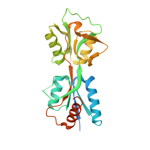
Structure of the effector-binding domain of the LysR-type transcription factor RovM from Yersinia pseudotuberculosis.

Quade, N.Dieckmann, M.Haffke, M.Heroven, A.K.Dersch, P.Heinz, D.W.

(2011) Acta Crystallogr D Biol Crystallogr 67: 81-90

  • DOI: https://doi.org/10.1107/S0907444910049681
  • Primary Citation Related Structures: 
    3ONM

  • PubMed Abstract: 

    In enteropathogenic Yersinia, the expression of several early-phase virulence factors such as invasin is tightly regulated in response to environmental cues. The responsible regulatory network is complex, involving several regulatory RNAs and proteins such as the LysR-type transcription regulator (LTTR) RovM. In this study, the crystal structure of the effector-binding domain (EBD) of RovM, the first LTTR protein described as being involved in virulence regulation, was determined at a resolution of 2.4 Å. Size-exclusion chromatography and comparison with structures of full-length LTTRs show that RovM is most likely to adopt a tetrameric arrangement with two distant DNA-binding domains (DBDs), causing the DNA to bend around it. Additionally, a cavity was detected in RovM which could bind small inducer molecules.


  • Organizational Affiliation
    • Department of Molecular Structural Biology, Helmholtz Centre for Infection Research, D-38124 Braunschweig, Germany.

Macromolecule Content 

  • Total Structure Weight: 52.32 kDa 
  • Atom Count: 2,857 
  • Modeled Residue Count: 370 
  • Deposited Residue Count: 476 
  • Unique protein chains: 1

Macromolecules

Find similar proteins by:|  3D Structure
Entity ID: 1
MoleculeChains  Sequence LengthOrganismDetailsImage
Transcriptional regulator LrhA
A, B
238Yersinia pseudotuberculosis IP 31758Mutation(s): 0 
Gene Names: lrhARovMYpsIP31758_1452

Experimental Data & Validation

Experimental Data

  • Method: X-RAY DIFFRACTION
  • Resolution: 2.40 Å
  • R-Value Free:  0.295 (Depositor), 0.278 (DCC) 
  • R-Value Work:  0.221 (Depositor), 0.214 (DCC) 
  • R-Value Observed: 0.224 (Depositor) 
Space Group: I 41 2 2
Unit Cell:
Length ( Å )Angle ( ˚ )
a = 69.468α = 90
b = 69.468β = 90
c = 351.216γ = 90
Software Package:
Software NamePurpose
SCALAdata scaling
SHELXphasing
REFMACrefinement
PDB_EXTRACTdata extraction
DNAdata collection
XDSdata reduction
SHELXDphasing

Structure Validation

View Full Validation Report


Entry History 

Deposition Data

Revision History  (Full details and data files)

  • Version 1.0: 2011-01-26
    Type: Initial release
  • Version 1.1: 2011-07-13
    Changes: Source and taxonomy, Version format compliance
  • Version 1.2: 2011-09-21
    Changes: Database references
  • Version 1.3: 2024-02-21
    Changes: Data collection, Database references