3OM3 | pdb_00003om3

Catalytic core subunits (I and II) of cytochrome C oxidase from Rhodobacter sphaeroides with K362M mutation in the reduced state


Experimental Data Snapshot

  • Method: X-RAY DIFFRACTION
  • Resolution: 2.60 Å
  • R-Value Free: 
    0.229 (Depositor), 0.234 (DCC) 
  • R-Value Work: 
    0.198 (Depositor) 
  • R-Value Observed: 
    0.199 (Depositor) 

wwPDB Validation 3D Report Full Report

Validation slider image for 3OM3

Ligand Structure Quality Assessment 


This is version 1.4 of the entry. See complete history

Literature

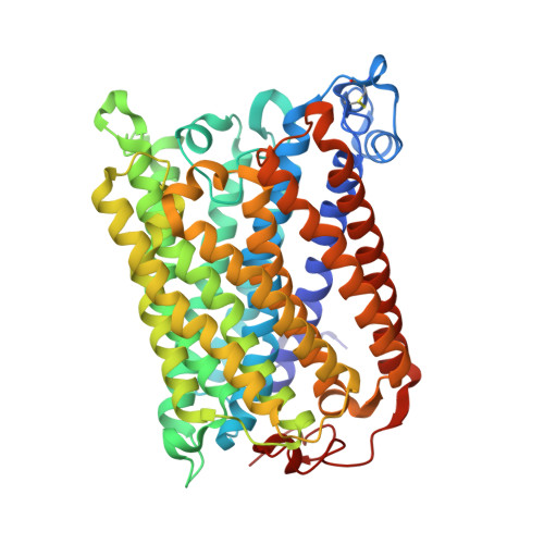
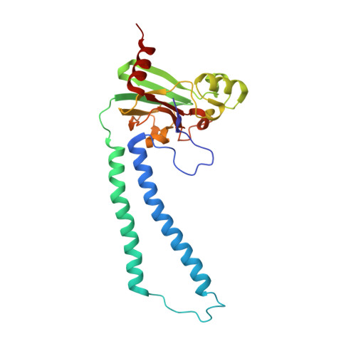
Crystallographic and online spectral evidence for role of conformational change and conserved water in cytochrome oxidase proton pump.

Liu, J.Qin, L.Ferguson-Miller, S.

(2011) Proc Natl Acad Sci U S A 108: 1284-1289

  • DOI: https://doi.org/10.1073/pnas.1012846108
  • Primary Citation Related Structures: 
    3OM3, 3OMA, 3OMI, 3OMN

  • PubMed Abstract: 

    Crystal structures in both oxidized and reduced forms are reported for two bacterial cytochrome c oxidase mutants that define the D and K proton paths, showing conformational change in response to reduction and the loss of strategic waters that can account for inhibition of proton transfer. In the oxidized state both mutants of the Rhodobacter sphaeroides enzyme, D132A and K362M, show overall structures similar to wild type, indicating no long-range effects of mutation. In the reduced state, the mutants show an altered conformation similar to that seen in reduced wild type, confirming this reproducible, reversible response to reduction. In the strongly inhibited D132A mutant, positions of residues and waters in the D pathway are unaffected except in the entry region close to the mutation, where a chloride ion replaces the missing carboxyl and a 2-Å shift in N207 results in loss of its associated water. In K362M, the methionine occupies the same position as the original lysine, but K362- and T359-associated waters in the wild-type structure are missing, likely accounting for the severe inhibition. Spectra of oxidized frozen crystals taken during X-ray radiation show metal center reduction, but indicate development of a strained configuration that only relaxes to a native form upon annealing. Resistance of the frozen crystal to structural change clarifies why the oxidized conformation is observable and supports the conclusion that the reduced conformation has functional significance. A mechanism is described that explains the conformational change and the incomplete response of the D-path mutant.


  • Organizational Affiliation
    • Department of Biochemistry and Molecular Biology, Michigan State University, East Lansing, MI 48824, USA.

Macromolecule Content 

  • Total Structure Weight: 187.81 kDa 
  • Atom Count: 13,203 
  • Modeled Residue Count: 1,578 
  • Deposited Residue Count: 1,582 
  • Unique protein chains: 2

Macromolecules

Find similar proteins by:|  3D Structure
Entity ID: 1
MoleculeChains  Sequence LengthOrganismDetailsImage
Cytochrome c oxidase, aa3 type, subunit I
A, C
535Cereibacter sphaeroides 2.4.1Mutation(s): 1 
Gene Names: coxICTADRHOS4_04590RSP_1877
EC: 1.9.3.1 (PDB Primary Data), 7.1.1.9 (UniProt)
Membrane Entity: Yes 
UniProt
Find proteins for Q3J5A7 (Cereibacter sphaeroides (strain ATCC 17023 / DSM 158 / JCM 6121 / CCUG 31486 / LMG 2827 / NBRC 12203 / NCIMB 8253 / ATH 2.4.1.))
Explore Q3J5A7 
Go to UniProtKB:  Q3J5A7
Entity Groups
Sequence Clusters30% Identity50% Identity70% Identity90% Identity95% Identity100% Identity
UniProt GroupQ3J5A7
Sequence Annotations
Expand
Reference Sequence
Find similar proteins by:|  3D Structure
Entity ID: 2
MoleculeChains  Sequence LengthOrganismDetailsImage
Cytochrome c oxidase subunit 2
B, D
256Cereibacter sphaeroides 2.4.1Mutation(s): 0 
Gene Names: coxIICTABCTACRHOS4_04060RSP_1826
EC: 1.9.3.1 (PDB Primary Data), 7.1.1.9 (UniProt)
Membrane Entity: Yes 
UniProt
Find proteins for Q3J5G0 (Cereibacter sphaeroides (strain ATCC 17023 / DSM 158 / JCM 6121 / CCUG 31486 / LMG 2827 / NBRC 12203 / NCIMB 8253 / ATH 2.4.1.))
Explore Q3J5G0 
Go to UniProtKB:  Q3J5G0
Entity Groups
Sequence Clusters30% Identity50% Identity70% Identity90% Identity95% Identity100% Identity
UniProt GroupQ3J5G0
Sequence Annotations
Expand
Reference Sequence

Small Molecules

Ligands 8 Unique
IDChains Name / Formula / InChI Key2D Diagram3D Interactions
HEA

Query on HEA


Download:Ideal Coordinates CCD File
DA [auth C],
EA [auth C],
J [auth A],
K [auth A]
HEME-A
C49 H56 Fe N4 O6
ZGGYGTCPXNDTRV-PRYGPKJJSA-L
DMU

Query on DMU


Download:Ideal Coordinates CCD File
AA [auth C]
BA [auth C]
CA [auth C]
E [auth A]
F [auth A]
AA [auth C],
BA [auth C],
CA [auth C],
E [auth A],
F [auth A],
KA [auth D],
LA [auth D],
Q [auth B],
R [auth B],
S [auth B],
T [auth B]
DECYL-BETA-D-MALTOPYRANOSIDE
C22 H42 O11
WOQQAWHSKSSAGF-WXFJLFHKSA-N
TRD

Query on TRD


Download:Ideal Coordinates CCD File
FA [auth C]
G [auth A]
GA [auth C]
H [auth A]
I [auth A]
FA [auth C],
G [auth A],
GA [auth C],
H [auth A],
I [auth A],
L [auth A],
M [auth A],
MA [auth D],
U [auth B]
TRIDECANE
C13 H28
IIYFAKIEWZDVMP-UHFFFAOYSA-N
HTH

Query on HTH


Download:Ideal Coordinates CCD File
V [auth B](2S,3R)-heptane-1,2,3-triol
C7 H16 O3
HXYCHJFUBNTKQR-RQJHMYQMSA-N
CD

Query on CD


Download:Ideal Coordinates CCD File
PA [auth D],
QA [auth D],
Y [auth B],
Z [auth B]
CADMIUM ION
Cd
WLZRMCYVCSSEQC-UHFFFAOYSA-N
CU1

Query on CU1


Download:Ideal Coordinates CCD File
HA [auth C]
N [auth A]
NA [auth D]
OA [auth D]
W [auth B]
HA [auth C],
N [auth A],
NA [auth D],
OA [auth D],
W [auth B],
X [auth B]
COPPER (I) ION
Cu
VMQMZMRVKUZKQL-UHFFFAOYSA-N
CA

Query on CA


Download:Ideal Coordinates CCD File
JA [auth C],
P [auth A]
CALCIUM ION
Ca
BHPQYMZQTOCNFJ-UHFFFAOYSA-N
MG

Query on MG


Download:Ideal Coordinates CCD File
IA [auth C],
O [auth A]
MAGNESIUM ION
Mg
JLVVSXFLKOJNIY-UHFFFAOYSA-N

Experimental Data & Validation

Experimental Data

  • Method: X-RAY DIFFRACTION
  • Resolution: 2.60 Å
  • R-Value Free:  0.229 (Depositor), 0.234 (DCC) 
  • R-Value Work:  0.198 (Depositor) 
  • R-Value Observed: 0.199 (Depositor) 
Space Group: P 21 21 21
Unit Cell:
Length ( Å )Angle ( ˚ )
a = 124.65α = 90
b = 132.365β = 90
c = 178.006γ = 90
Software Package:
Software NamePurpose
DENZOdata reduction
SCALEPACKdata scaling
REFMACrefinement
PDB_EXTRACTdata extraction
MD2data collection
HKL-2000data reduction
HKL-2000data scaling
CNSphasing

Structure Validation

View Full Validation Report


Ligand Structure Quality Assessment 


Entry History 

Deposition Data

Revision History  (Full details and data files)

  • Version 1.0: 2011-02-02
    Type: Initial release
  • Version 1.1: 2011-07-13
    Changes: Version format compliance
  • Version 1.2: 2012-05-02
    Changes: Structure summary
  • Version 1.3: 2017-11-08
    Changes: Refinement description
  • Version 1.4: 2024-11-06
    Changes: Data collection, Database references, Derived calculations, Structure summary