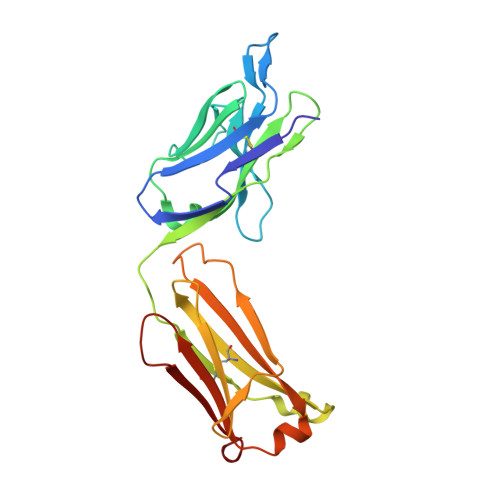
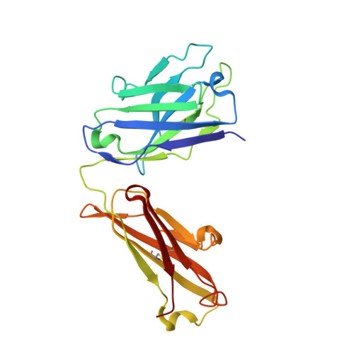
3OKL | pdb_00003okl

Crystal structure of S25-39 in complex with Kdo(2.8)Kdo


Experimental Data Snapshot

  • Method: X-RAY DIFFRACTION
  • Resolution: 1.80 Å
  • R-Value Free: 
    0.240 (Depositor), 0.231 (DCC) 
  • R-Value Work: 
    0.191 (Depositor), 0.183 (DCC) 
  • R-Value Observed: 
    0.193 (Depositor) 

wwPDB Validation 3D Report Full Report

Validation slider image for 3OKL

This is version 2.1 of the entry. See complete history. 

Literature

A Common NH53K Mutation in the Combining Site of Antibodies Raised against Chlamydial LPS Glycoconjugates Significantly Increases Avidity.

Blackler, R.J., Brooks, C.L., Evans, D.W., Brade, L., Kosma, P., Brade, H., Evans, S.V.

(2011) Biochemistry 50: 3357-3368

  • DOI: https://doi.org/10.1021/bi101886v
  • Primary Citation Related Structures: 
    3OKD, 3OKE, 3OKK, 3OKL, 3OKM, 3OKN, 3OKO

  • PubMed Abstract: 

    The crystal structures of the antigen-binding fragment of the murine monoclonal antibody (mAb) S25-39 in the presence of several antigens representing chlamydial lipopolysaccharide (LPS) epitopes based on the bacterial sugar 3-deoxy-α-D-manno-oct-2-ulosonic acid (Kdo) have been determined at resolutions from 2.4 to 1.8 Å. The antigen-binding site of this antibody differs from the well-characterized antibody S25-2 by a single mutation away from the germline of asparagine H53 to lysine, yet this one mutation results in a significant increase in avidity across a range of antigens. A comparison of the two antibody structures reveals that the mutated Lys H53 forms additional hydrogen bonds and/or charged-residue interactions with the second Kdo residue of every antigen having two or more carbohydrate residues. Significantly, the NH53K mutation results from a single nucleotide substitution in the germline sequence common among a panel of antibodies raised against glycoconjugates containing carbohydrate epitopes of chlamydial LPS. Like S25-2, S25-39 displays significant induced fit of complementarity determining region (CDR) H3 upon antigen binding, with the unliganded structure possessing a conformation distinct from those reported earlier for S25-2. The four different observed conformations for CDR H3 suggest that this CDR has evolved to exploit the recognition potential of a flexible loop while minimizing the associated entropic penalties of binding by adopting a limited number of ordered conformations in the unliganded state. These observations reveal strategies evolved to balance adaptability and specificity in the germline antibody response to carbohydrate antigens.


  • Organizational Affiliation: 
    • Department of Biochemistry and Microbiology, University of Victoria , Victoria, BC V8P 3P6, Canada.

Macromolecule Content 

  • Total Structure Weight: 48.77 kDa 
  • Atom Count: 3,961 
  • Modeled Residue Count: 441 
  • Deposited Residue Count: 441 
  • Unique protein chains: 2

Macromolecules

Find similar proteins by:|  3D Structure
Entity ID: 1
MoleculeChains  Sequence LengthOrganismDetailsImage
S25-39 Fab (IgG1k) light chain219Mus musculusMutation(s): 0 
Entity Groups
Sequence Clusters30% Identity50% Identity70% Identity90% Identity95% Identity100% Identity
Sequence Annotations
Expand
Reference Sequence
Find similar proteins by:|  3D Structure
Entity ID: 2
MoleculeChains  Sequence LengthOrganismDetailsImage
S25-39 Fab (IgG1k) heavy chain222Mus musculusMutation(s): 0 
Entity Groups
Sequence Clusters30% Identity50% Identity70% Identity90% Identity95% Identity100% Identity
Sequence Annotations
Expand
Reference Sequence

Oligosaccharides

Help  
Entity ID: 3
MoleculeChains Length2D Diagram GlycosylationD Interactions
3-deoxy-alpha-D-manno-oct-2-ulopyranosonic acid-(2-8)-prop-2-en-1-yl 3-deoxy-alpha-D-manno-oct-2-ulopyranosidonic acid
C
2N/AN/A
Glycosylation Resources
GlyTouCan: G82878QT
GlyCosmos: G82878QT

Small Molecules

Ligands 1 Unique
IDChains Name / Formula / InChI Key2D Diagram3D Interactions
ZN

Query on ZN


Download:Ideal Coordinates CCD File
D [auth B]ZINC ION
Zn
PTFCDOFLOPIGGS-UHFFFAOYSA-N

Experimental Data & Validation

Experimental Data

  • Method: X-RAY DIFFRACTION
  • Resolution: 1.80 Å
  • R-Value Free:  0.240 (Depositor), 0.231 (DCC) 
  • R-Value Work:  0.191 (Depositor), 0.183 (DCC) 
  • R-Value Observed: 0.193 (Depositor) 
Space Group: P 21 21 21
Unit Cell:
Length ( Å )Angle ( ˚ )
a = 45.525α = 90
b = 81.657β = 90
c = 131.868γ = 90
Software Package:
Software NamePurpose
d*TREKdata scaling
PHASERphasing
PHENIXrefinement
PDB_EXTRACTdata extraction

Structure Validation

View Full Validation Report


Entry History 

Deposition Data

Revision History  (Full details and data files)

  • Version 1.0: 2011-04-06
    Type: Initial release
  • Version 1.1: 2011-07-13
    Changes: Version format compliance
  • Version 1.2: 2018-03-07
    Changes: Data collection
  • Version 2.0: 2020-07-29
    Type: Remediation
    Reason: Carbohydrate remediation
    Changes: Atomic model, Data collection, Derived calculations, Structure summary
  • Version 2.1: 2024-10-16
    Changes: Data collection, Database references, Structure summary