


Crystal structure of MDD in complex with Dihydroxyacetone
Zhao, H.-T., El-Kabbani, O., Hara, A.To be published.
Experimental Data Snapshot
Starting Model: experimental
View more details
wwPDB Validation 3D Report Full Report

Entity ID: 1 | |||||
|---|---|---|---|---|---|
| Molecule | Chains | Sequence Length | Organism | Details | Image |
| Trans-1,2-dihydrobenzene-1,2-diol dehydrogenase | A [auth X] | 334 | Macaca fascicularis | Mutation(s): 0 Gene Names: DHDH, 2DD EC: 1.3.1.20 (PDB Primary Data), 1.1.1.179 (PDB Primary Data), 1.1.1.119 (UniProt), 1.1.1.115 (UniProt), 1.1.1 (UniProt), 1.1.1.120 (UniProt) | 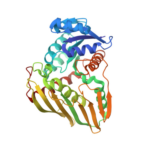 |
UniProt | |||||
Entity Groups | |||||
| Sequence Clusters | 30% Identity50% Identity70% Identity90% Identity95% Identity100% Identity | ||||
| UniProt Group | Q9TQS6 | ||||
Sequence AnnotationsExpand | |||||
Reference Sequence | |||||
| Ligands 3 Unique | |||||
|---|---|---|---|---|---|
| ID | Chains | Name / Formula / InChI Key | 2D Diagram | 3D Interactions | |
| SO4 Download:Ideal Coordinates CCD File | B [auth X] C [auth X] D [auth X] E [auth X] F [auth X] | SULFATE ION O4 S QAOWNCQODCNURD-UHFFFAOYSA-L |  | ||
| 2HA Download:Ideal Coordinates CCD File | K [auth X] | Dihydroxyacetone C3 H6 O3 RXKJFZQQPQGTFL-UHFFFAOYSA-N |  | ||
| BME Download:Ideal Coordinates CCD File | J [auth X] | BETA-MERCAPTOETHANOL C2 H6 O S DGVVWUTYPXICAM-UHFFFAOYSA-N |  | ||
| Length ( Å ) | Angle ( ˚ ) |
|---|---|
| a = 122.446 | α = 90 |
| b = 122.446 | β = 90 |
| c = 121.388 | γ = 120 |
| Software Name | Purpose |
|---|---|
| HKL-2000 | data collection |
| MOLREP | phasing |
| REFMAC | refinement |
| HKL-2000 | data reduction |
| SCALEPACK | data scaling |


