3OHG | pdb_00003ohg

Crystal structure of a protein with unknown function from DUF2233 family (BACOVA_00430) from Bacteroides ovatus at 1.80 A resolution


Experimental Data Snapshot

  • Method: X-RAY DIFFRACTION
  • Resolution: 1.80 Å
  • R-Value Free: 
    0.151 (Depositor), 0.163 (DCC) 
  • R-Value Work: 
    0.129 (Depositor), 0.142 (DCC) 
  • R-Value Observed: 
    0.130 (Depositor) 

wwPDB Validation 3D Report Full Report

Validation slider image for 3OHG

This is version 1.5 of the entry. See complete history

Literature

Structure and function of the DUF2233 domain in bacteria and in the human mannose 6-phosphate uncovering enzyme.

Das, D.Lee, W.S.Grant, J.C.Chiu, H.J.Farr, C.L.Vance, J.Klock, H.E.Knuth, M.W.Miller, M.D.Elsliger, M.A.Deacon, A.M.Godzik, A.Lesley, S.A.Kornfeld, S.Wilson, I.A.

(2013) J Biological Chem 288: 16789-16799

  • DOI: https://doi.org/10.1074/jbc.M112.434977
  • Primary Citation Related Structures: 
    3OHG

  • PubMed Abstract: 

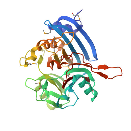
    DUF2233, a domain of unknown function (DUF), is present in many bacterial and several viral proteins and was also identified in the mammalian transmembrane glycoprotein N-acetylglucosamine-1-phosphodiester α-N-acetylglucosaminidase ("uncovering enzyme" (UCE)). We report the crystal structure of BACOVA_00430, a 315-residue protein from the human gut bacterium Bacteroides ovatus that is the first structural representative of the DUF2233 protein family. A notable feature of this structure is the presence of a surface cavity that is populated by residues that are highly conserved across the entire family. The crystal structure was used to model the luminal portion of human UCE (hUCE), which is involved in targeting of lysosomal enzymes. Mutational analysis of several residues in a highly conserved surface cavity of hUCE revealed that they are essential for function. The bacterial enzyme (BACOVA_00430) has ∼1% of the catalytic activity of hUCE toward the substrate GlcNAc-P-mannose, the precursor of the Man-6-P lysosomal targeting signal. GlcNAc-1-P is a poor substrate for both enzymes. We conclude that, for at least a subset of proteins in this family, DUF2233 functions as a phosphodiester glycosidase.


  • Organizational Affiliation
    • Joint Center for Structural Genomics, Menlo Park, California 94025; Stanford Synchrotron Radiation Lightsource, SLAC National Accelerator Laboratory, Menlo Park, California 94025.

Macromolecule Content 

  • Total Structure Weight: 32.82 kDa 
  • Atom Count: 2,815 
  • Modeled Residue Count: 285 
  • Deposited Residue Count: 285 
  • Unique protein chains: 1

Macromolecules

Find similar proteins by:|  3D Structure
Entity ID: 1
MoleculeChains  Sequence LengthOrganismDetailsImage
Uncharacterized protein from DUF2233 family285Bacteroides ovatus ATCC 8483Mutation(s): 0 
Gene Names: BACOVA_00430

Small Molecules

Ligands 3 Unique
IDChains Name / Formula / InChI Key2D Diagram3D Interactions
SO4

Query on SO4


Download:Ideal Coordinates CCD File
C [auth A],
D [auth A]
SULFATE ION
O4 S
QAOWNCQODCNURD-UHFFFAOYSA-L
EDO

Query on EDO


Download:Ideal Coordinates CCD File
E [auth A]
F [auth A]
G [auth A]
H [auth A]
I [auth A]
E [auth A],
F [auth A],
G [auth A],
H [auth A],
I [auth A],
J [auth A],
K [auth A],
L [auth A],
M [auth A],
N [auth A],
O [auth A],
P [auth A],
Q [auth A],
R [auth A],
S [auth A],
T [auth A],
U [auth A]
1,2-ETHANEDIOL
C2 H6 O2
LYCAIKOWRPUZTN-UHFFFAOYSA-N
CL

Query on CL


Download:Ideal Coordinates CCD File
B [auth A]CHLORIDE ION
Cl
VEXZGXHMUGYJMC-UHFFFAOYSA-M
Modified Residues  1 Unique
IDChains TypeFormula2D DiagramParent
MSE
Query on MSE
A
L-PEPTIDE LINKINGC5 H11 N O2 SeMET

Experimental Data & Validation

Experimental Data

  • Method: X-RAY DIFFRACTION
  • Resolution: 1.80 Å
  • R-Value Free:  0.151 (Depositor), 0.163 (DCC) 
  • R-Value Work:  0.129 (Depositor), 0.142 (DCC) 
  • R-Value Observed: 0.130 (Depositor) 
Space Group: P 61
Unit Cell:
Length ( Å )Angle ( ˚ )
a = 96.964α = 90
b = 96.964β = 90
c = 91.113γ = 120
Software Package:
Software NamePurpose
SHELXphasing
REFMACrefinement
SCALAdata scaling
PDB_EXTRACTdata extraction
MOSFLMdata reduction
SHELXDphasing
autoSHARPphasing

Structure Validation

View Full Validation Report


Entry History 

Deposition Data

Revision History  (Full details and data files)

  • Version 1.0: 2010-09-08
    Type: Initial release
  • Version 1.1: 2011-07-13
    Changes: Version format compliance
  • Version 1.2: 2011-07-20
    Changes: Structure summary
  • Version 1.3: 2014-09-24
    Changes: Database references
  • Version 1.4: 2023-02-01
    Changes: Database references, Derived calculations
  • Version 1.5: 2024-11-20
    Changes: Data collection, Structure summary