3OHF | pdb_00003ohf

Crystal structure of beta-site app-cleaving enzyme 1 (BACE-WT) complex with bms-655295 aka n~3~-((1s,2r)-1- benzyl-2-hydroxy-3-((3-methoxybenzyl)amino)propyl)-n~1~, n~1~-dibutyl-1h-indole-1,3-dicarboxamide


Experimental Data Snapshot

  • Method: X-RAY DIFFRACTION
  • Resolution: 2.10 Å
  • R-Value Free: 
    0.248 (Depositor) 
  • R-Value Work: 
    0.203 (Depositor) 
  • R-Value Observed: 
    0.205 (Depositor) 

wwPDB Validation 3D Report Full Report

Validation slider image for 3OHF

Ligand Structure Quality Assessment 


This is version 1.3 of the entry. See complete history

Literature

Synthesis and SAR of indole-and 7-azaindole-1,3-dicarboxamide hydroxyethylamine inhibitors of BACE-1.

Marcin, L.R.Higgins, M.A.Zusi, F.C.Zhang, Y.Dee, M.F.Parker, M.F.Muckelbauer, J.K.Camac, D.M.Morin, P.E.Ramamurthy, V.Tebben, A.J.Lentz, K.A.Grace, J.E.Marcinkeviciene, J.A.Kopcho, L.M.Burton, C.R.Barten, D.M.Toyn, J.H.Meredith, J.E.Albright, C.F.Bronson, J.J.Macor, J.E.Thompson, L.A.

(2011) Bioorg Med Chem Lett 21: 537-541

  • DOI: https://doi.org/10.1016/j.bmcl.2010.10.079
  • Primary Citation Related Structures: 
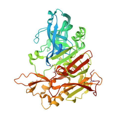
    3OHF, 3OHH

  • PubMed Abstract: 

    Heterocyclic replacement of the isophthalamide phenyl ring in hydroxyethylamine (HEA) BACE-1 inhibitors was explored. A variety of indole-1,3-dicarboxamide HEAs exhibited potent BACE-1 enzyme inhibition, but displayed poor cellular activity. Improvements in cellular activity and aspartic protease selectivity were observed for 7-azaindole-1,3-dicarboxamide HEAs. A methylprolinol-bearing derivative (10n) demonstrated robust reductions in rat plasma Aβ levels, but did not lower rat brain Aβ due to poor central exposure. The same analog exhibited a high efflux ratio in a bidirectional Caco-2 assay and was likely a substrate of the efflux transporter P-glycoprotein. X-ray crystal structures are reported for two indole HEAs in complex with BACE-1.


  • Organizational Affiliation
    • Bristol-Myers Squibb, Research and Development, 5 Research Parkway, Wallingford, CT 06492-7660, USA. lawrence.marcin@bms.com

Macromolecule Content 

  • Total Structure Weight: 102.08 kDa 
  • Atom Count: 6,977 
  • Modeled Residue Count: 774 
  • Deposited Residue Count: 910 
  • Unique protein chains: 1

Macromolecules

Find similar proteins by:|  3D Structure
Entity ID: 1
MoleculeChains  Sequence LengthOrganismDetailsImage
Beta-secretase 1
A, B
455Homo sapiensMutation(s): 0 
Gene Names: BACEBACE1KIAA1149
EC: 3.4.23.46
UniProt & NIH Common Fund Data Resources
Find proteins for P56817 (Homo sapiens)
Explore P56817 
Go to UniProtKB:  P56817
PHAROS:  P56817
GTEx:  ENSG00000186318 
Entity Groups
Sequence Clusters30% Identity50% Identity70% Identity90% Identity95% Identity100% Identity
UniProt GroupP56817
Sequence Annotations
Expand
Reference Sequence

Small Molecules

Ligands 3 Unique
IDChains Name / Formula / InChI Key2D Diagram3D Interactions
3HF

Query on 3HF


Download:Ideal Coordinates CCD File
C [auth A],
F [auth B]
N~3~-{(1S,2R)-1-benzyl-2-hydroxy-3-[(3-methoxybenzyl)amino]propyl}-N~1~,N~1~-dibutyl-1H-indole-1,3-dicarboxamide
C36 H46 N4 O4
JPMPCSZOVMUPRZ-UZNNEEJFSA-N
IOD

Query on IOD


Download:Ideal Coordinates CCD File
D [auth A],
G [auth B]
IODIDE ION
I
XMBWDFGMSWQBCA-UHFFFAOYSA-M
GOL

Query on GOL


Download:Ideal Coordinates CCD File
E [auth A],
H [auth B]
GLYCEROL
C3 H8 O3
PEDCQBHIVMGVHV-UHFFFAOYSA-N

Experimental Data & Validation

Experimental Data

  • Method: X-RAY DIFFRACTION
  • Resolution: 2.10 Å
  • R-Value Free:  0.248 (Depositor) 
  • R-Value Work:  0.203 (Depositor) 
  • R-Value Observed: 0.205 (Depositor) 
Space Group: P 21 21 2
Unit Cell:
Length ( Å )Angle ( ˚ )
a = 88.982α = 90
b = 130.233β = 90
c = 86.596γ = 90
Software Package:
Software NamePurpose
SCALEPACKdata scaling
REFMACrefinement
PDB_EXTRACTdata extraction
HKL-2000data collection
HKL-2000data reduction
HKL-2000data scaling

Structure Validation

View Full Validation Report


Ligand Structure Quality Assessment 


Entry History 

Deposition Data

Revision History  (Full details and data files)

  • Version 1.0: 2011-04-06
    Type: Initial release
  • Version 1.1: 2011-07-13
    Changes: Version format compliance
  • Version 1.2: 2017-03-01
    Changes: Database references
  • Version 1.3: 2024-10-16
    Changes: Data collection, Database references, Derived calculations, Structure summary