3OH6 | pdb_00003oh6

AlkA Undamaged DNA Complex: Interrogation of a C:G base pair


Experimental Data Snapshot

  • Method: X-RAY DIFFRACTION
  • Resolution: 2.89 Å
  • R-Value Free: 
    0.269 (Depositor), 0.260 (DCC) 
  • R-Value Work: 
    0.227 (Depositor), 0.216 (DCC) 
  • R-Value Observed: 
    0.229 (Depositor) 

Starting Model: experimental
View more details

wwPDB Validation 3D Report Full Report

Validation slider image for 3OH6

This is version 1.2 of the entry. See complete history

Literature

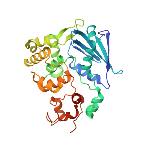
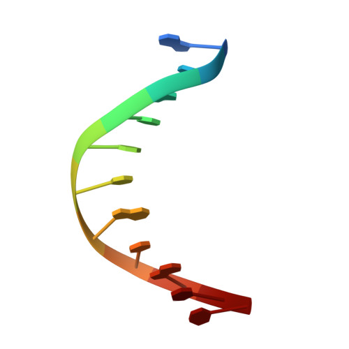
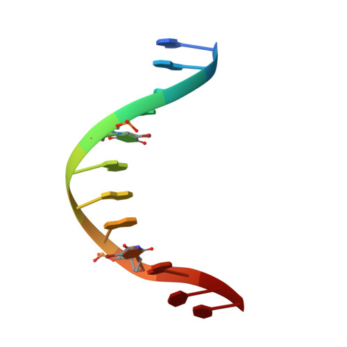
Structure of Escherichia coli AlkA in Complex with Undamaged DNA.

Bowman, B.R.Lee, S.Wang, S.Verdine, G.L.

(2010) J Biological Chem 285: 35783-35791

  • DOI: https://doi.org/10.1074/jbc.M110.155663
  • Primary Citation Related Structures: 
    3OGD, 3OH6, 3OH9

  • PubMed Abstract: 

    Because DNA damage is so rare, DNA glycosylases interact for the most part with undamaged DNA. Whereas the structural basis for recognition of DNA lesions by glycosylases has been studied extensively, less is known about the nature of the interaction between these proteins and undamaged DNA. Here we report the crystal structures of the DNA glycosylase AlkA in complex with undamaged DNA. The structures revealed a recognition mode in which the DNA is nearly straight, with no amino acid side chains inserted into the duplex, and the target base pair is fully intrahelical. A comparison of the present structures with that of AlkA recognizing an extrahelical lesion revealed conformational changes in both the DNA and protein as the glycosylase transitions from the interrogation of undamaged DNA to catalysis of nucleobase excision. Modeling studies with the cytotoxic lesion 3-methyladenine and accompanying biochemical experiments suggested that AlkA actively interrogates the minor groove of the DNA while probing for the presence of lesions.


  • Organizational Affiliation
    • Department of Stem Cell and Regenerative Biology, Harvard University, Cambridge, Massachusetts 02138, USA.

Macromolecule Content 

  • Total Structure Weight: 39.21 kDa 
  • Atom Count: 2,718 
  • Modeled Residue Count: 311 
  • Deposited Residue Count: 312 
  • Unique protein chains: 1
  • Unique nucleic acid chains: 2

Macromolecules


Find similar proteins by:|  3D Structure
Entity ID: 1
MoleculeChains  Sequence LengthOrganismDetailsImage
DNA-3-methyladenine glycosylase 2289Escherichia coli K-12Mutation(s): 1 
Gene Names: aidAalkAb2068JW2053
EC: 3.2.2.21
UniProt
Find proteins for P04395 (Escherichia coli (strain K12))
Explore P04395 
Go to UniProtKB:  P04395
Entity Groups
Sequence Clusters30% Identity50% Identity70% Identity90% Identity95% Identity100% Identity
UniProt GroupP04395
Sequence Annotations
Expand
Reference Sequence
Find similar nucleic acids by:  Sequence
Entity ID: 2
MoleculeChains LengthOrganismImage
5'-D(*GP*CP*AP*TP*TP*CP*AP*TP*GP*TP*C)-3'11N/A
Sequence Annotations
Expand
Reference Sequence
Find similar nucleic acids by:  Sequence
Entity ID: 3
MoleculeChains LengthOrganismImage
5'-D(*GP*AP*CP*AP*(BRU)P*GP*AP*AP*(BRU)P*GP*CP*C)-3'12N/A
Sequence Annotations
Expand
Reference Sequence

Experimental Data & Validation

Experimental Data

  • Method: X-RAY DIFFRACTION
  • Resolution: 2.89 Å
  • R-Value Free:  0.269 (Depositor), 0.260 (DCC) 
  • R-Value Work:  0.227 (Depositor), 0.216 (DCC) 
  • R-Value Observed: 0.229 (Depositor) 
Space Group: I 41 2 2
Unit Cell:
Length ( Å )Angle ( ˚ )
a = 140.3α = 90
b = 140.3β = 90
c = 94.8γ = 90
Software Package:
Software NamePurpose
SCALEPACKdata scaling
PHENIXrefinement
PDB_EXTRACTdata extraction
ADSCdata collection
HKL-2000data reduction
CNSphasing

Structure Validation

View Full Validation Report


Entry History 

Deposition Data

Revision History  (Full details and data files)

  • Version 1.0: 2010-09-15
    Type: Initial release
  • Version 1.1: 2011-07-13
    Changes: Version format compliance
  • Version 1.2: 2023-09-06
    Changes: Data collection, Database references, Derived calculations, Refinement description