3OG5 | pdb_00003og5

Crystal Structure of BamA POTRA45 tandem


Experimental Data Snapshot

  • Method: X-RAY DIFFRACTION
  • Resolution: 2.69 Å
  • R-Value Free: 
    0.260 (Depositor), 0.256 (DCC) 
  • R-Value Work: 
    0.211 (Depositor), 0.213 (DCC) 
  • R-Value Observed: 
    0.216 (Depositor) 

wwPDB Validation 3D Report Full Report

Validation slider image for 3OG5

This is version 1.2 of the entry. See complete history

Literature

Structure and Flexibility of the Complete Periplasmic Domain of BamA: The Protein Insertion Machine of the Outer Membrane

Gatzeva-Topalova, P.Z.Warner, L.R.Pardi, A.Sousa, M.C.

(2010) Structure 18: 1492-1501

  • DOI: https://doi.org/10.1016/j.str.2010.08.012
  • Primary Citation Related Structures: 
    3OG5

  • PubMed Abstract: 

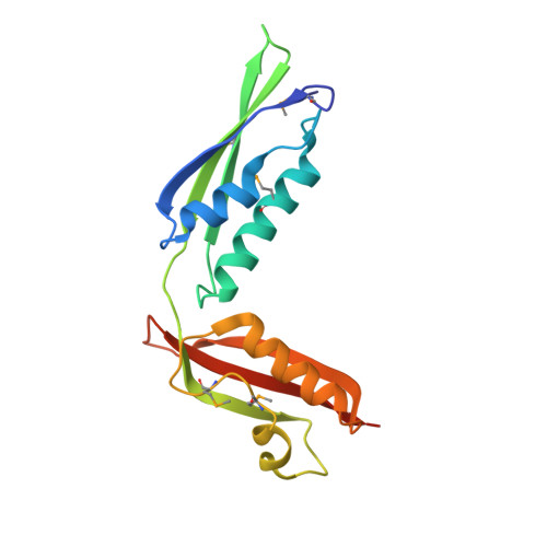
    Folding and insertion of β-barrel outer membrane proteins (OMPs) is essential for Gram-negative bacteria. This process is mediated by the multiprotein complex BAM, composed of the essential β-barrel OMP BamA and four lipoproteins (BamBCDE). The periplasmic domain of BamA is key for its function and contains five "polypeptide transport-associated" (POTRA) repeats. Here, we report the crystal structure of the POTRA4-5 tandem, containing the essential for BAM complex formation and cell viability POTRA5. The domain orientation observed in the crystal is validated by solution NMR and SAXS. Using previously determined structures of BamA POTRA1-4, we present a spliced model of the entire BamA periplasmic domain validated by SAXS. Solution scattering shows that conformational flexibility between POTRA2 and 3 gives rise to compact and extended conformations. The length of BamA in its extended conformation suggests that the protein may bridge the inner and outer membranes across the periplasmic space.


  • Organizational Affiliation
    • Department of Chemistry and Biochemistry, University of Colorado at Boulder, Boulder, CO 80309, USA.

Macromolecule Content 

  • Total Structure Weight: 37.87 kDa 
  • Atom Count: 2,351 
  • Modeled Residue Count: 296 
  • Deposited Residue Count: 332 
  • Unique protein chains: 1

Macromolecules

Find similar proteins by:|  3D Structure
Entity ID: 1
MoleculeChains  Sequence LengthOrganismDetailsImage
Outer membrane protein assembly complex, YaeT protein
A, B
166Escherichia coli K-12Mutation(s): 0 
Gene Names: yaeT
Membrane Entity: Yes 
UniProt
Find proteins for P0A940 (Escherichia coli (strain K12))
Explore P0A940 
Go to UniProtKB:  P0A940
Entity Groups
Sequence Clusters30% Identity50% Identity70% Identity90% Identity95% Identity100% Identity
UniProt GroupP0A940
Sequence Annotations
Expand
Reference Sequence

Small Molecules

Modified Residues  1 Unique
IDChains TypeFormula2D DiagramParent
MSE
Query on MSE
A, B
L-PEPTIDE LINKINGC5 H11 N O2 SeMET

Experimental Data & Validation

Experimental Data

  • Method: X-RAY DIFFRACTION
  • Resolution: 2.69 Å
  • R-Value Free:  0.260 (Depositor), 0.256 (DCC) 
  • R-Value Work:  0.211 (Depositor), 0.213 (DCC) 
  • R-Value Observed: 0.216 (Depositor) 
Space Group: P 32 2 1
Unit Cell:
Length ( Å )Angle ( ˚ )
a = 118.492α = 90
b = 118.492β = 90
c = 69.992γ = 120
Software Package:
Software NamePurpose
SCALEPACKdata scaling
PHENIXrefinement
PDB_EXTRACTdata extraction
HKL-2000data collection
HKL-2000data reduction
MOLREPphasing
PHENIXphasing

Structure Validation

View Full Validation Report


Entry History 

Deposition Data

Revision History  (Full details and data files)

  • Version 1.0: 2010-11-17
    Type: Initial release
  • Version 1.1: 2011-07-13
    Changes: Version format compliance
  • Version 1.2: 2024-10-16
    Changes: Data collection, Database references, Derived calculations, Structure summary