3OEK | pdb_00003oek

Crystal structure of GluN2D ligand-binding core in complex with L-aspartate


Experimental Data Snapshot

  • Method: X-RAY DIFFRACTION
  • Resolution: 1.90 Å
  • R-Value Free: 
    0.227 (Depositor), 0.227 (DCC) 
  • R-Value Work: 
    0.189 (Depositor), 0.188 (DCC) 
  • R-Value Observed: 
    0.191 (Depositor) 

wwPDB Validation 3D Report Full Report

Validation slider image for 3OEK

This is version 1.3 of the entry. See complete history

Literature

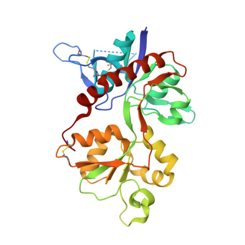
Ligand-specific deactivation time course of GluN1/GluN2D NMDA receptors.

Vance, K.M.Simorowski, N.Traynelis, S.F.Furukawa, H.

(2011) Nat Commun 2: 294-294

  • DOI: https://doi.org/10.1038/ncomms1295
  • Primary Citation Related Structures: 
    3OEK, 3OEL, 3OEM, 3OEN

  • PubMed Abstract: 

    N-methyl-D-aspartate (NMDA) receptors belong to the family of ionotropic glutamate receptors that mediate a majority of excitatory synaptic transmission. One unique property of GluN1/GluN2D NMDA receptors is an unusually prolonged deactivation time course following the removal of L-glutamate. Here we show, using x-ray crystallography and electrophysiology, that the deactivation time course of GluN1/GluN2D receptors is influenced by the conformational variability of the ligand-binding domain (LBD) as well as the structure of the activating ligand. L-glutamate and L-CCG-IV induce significantly slower deactivation time courses compared with other agonists. Crystal structures of the isolated GluN2D LBD in complex with various ligands reveal that the binding of L-glutamate induces a unique conformation at the backside of the ligand-binding site in proximity to the region at which the transmembrane domain would be located in the intact receptors. These data suggest that the activity of the GluN1/GluN2D NMDA receptor is controlled distinctively by the endogenous neurotransmitter L-glutamate.


  • Organizational Affiliation
    • Department of Pharmacology, Emory University School of Medicine, Rollins Research Center, 1510 Clifton Road, Atlanta, Georgia 30322-3090, USA.

Macromolecule Content 

  • Total Structure Weight: 32.17 kDa 
  • Atom Count: 2,381 
  • Modeled Residue Count: 270 
  • Deposited Residue Count: 286 
  • Unique protein chains: 1

Macromolecules

Find similar proteins by:|  3D Structure
Entity ID: 1
MoleculeChains  Sequence LengthOrganismDetailsImage
Glutamate [NMDA] receptor subunit epsilon-4286Rattus norvegicusMutation(s): 0 
Gene Names: GluN2DGrin2d
UniProt
Find proteins for Q62645 (Rattus norvegicus)
Explore Q62645 
Go to UniProtKB:  Q62645
Entity Groups
Sequence Clusters30% Identity50% Identity70% Identity90% Identity95% Identity100% Identity
UniProt GroupQ62645
Sequence Annotations
Expand
Reference Sequence

Small Molecules

Ligands 1 Unique
IDChains Name / Formula / InChI Key2D Diagram3D Interactions
ASP

Query on ASP


Download:Ideal Coordinates CCD File
B [auth A]ASPARTIC ACID
C4 H7 N O4
CKLJMWTZIZZHCS-REOHCLBHSA-N

Experimental Data & Validation

Experimental Data

  • Method: X-RAY DIFFRACTION
  • Resolution: 1.90 Å
  • R-Value Free:  0.227 (Depositor), 0.227 (DCC) 
  • R-Value Work:  0.189 (Depositor), 0.188 (DCC) 
  • R-Value Observed: 0.191 (Depositor) 
Space Group: C 2 2 21
Unit Cell:
Length ( Å )Angle ( ˚ )
a = 59.728α = 90
b = 114.062β = 90
c = 94.928γ = 90
Software Package:
Software NamePurpose
PHENIXrefinement

Structure Validation

View Full Validation Report


Entry History 

Deposition Data

Revision History  (Full details and data files)

  • Version 1.0: 2011-05-11
    Type: Initial release
  • Version 1.1: 2011-07-13
    Changes: Version format compliance
  • Version 1.2: 2017-08-23
    Changes: Source and taxonomy
  • Version 1.3: 2024-10-09
    Changes: Data collection, Database references, Derived calculations, Structure summary