3OEI | pdb_00003oei

Crystal structure of Mycobacterium tuberculosis RelJK (Rv3357-Rv3358-RelBE3)


Experimental Data Snapshot

  • Method: X-RAY DIFFRACTION
  • Resolution: 2.15 Å
  • R-Value Free: 
    0.256 (Depositor), 0.249 (DCC) 
  • R-Value Work: 
    0.203 (Depositor), 0.202 (DCC) 
  • R-Value Observed: 
    0.206 (Depositor) 

Starting Model: experimental
View more details

wwPDB Validation 3D Report Full Report

Validation slider image for 3OEI

This is version 1.4 of the entry. See complete history

Literature

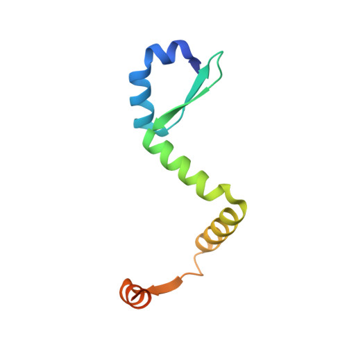
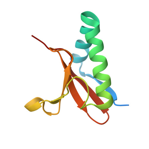
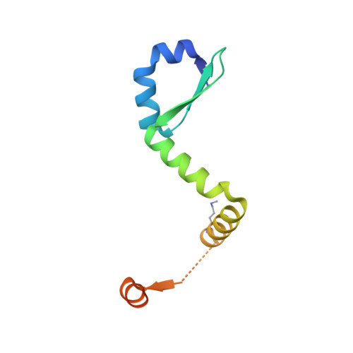
Comparative proteomics identifies the cell-associated lethality of M. tuberculosis RelBE-like toxin-antitoxin complexes.

Miallau, L.Jain, P.Arbing, M.A.Cascio, D.Phan, T.Ahn, C.J.Chan, S.Chernishof, I.Maxson, M.Chiang, J.Jacobs Jr., W.R.Eisenberg, D.S.

(2013) Structure 21: 627-637

  • DOI: https://doi.org/10.1016/j.str.2013.02.008
  • Primary Citation Related Structures: 
    3G5O, 3OEI

  • PubMed Abstract: 

    The Mycobacterium tuberculosis (Mtb) genome encodes approximately 90 toxin-antitoxin protein complexes, including three RelBE family members, which are believed to play a major role in bacterial fitness and pathogenicity. We have determined the crystal structures of Mtb RelBE-2 and RelBE-3, and the structures reveal homologous heterotetramers. Our structures suggest RelE-2, and by extension the closely related RelE-1, use a different catalytic mechanism than RelE-3, because our analysis of the RelE-2 structure predicts additional amino acid residues that are likely to be functionally significant and are missing from analogous positions in the RelE-3 structure. Toxicity assays corroborate our structural findings; overexpression of RelE-3, whose active site is more similar to Escherichia coli YoeB, has limited consequences on bacterial growth, whereas RelE-1 and RelE-2 overexpression results in acute toxicity. Moreover, RelE-2 overexpression results in an elongated cell phenotype in Mycobacterium smegmatis and protects M. tuberculosis against antibiotics, suggesting a different functional role for RelE-2.


  • Organizational Affiliation
    • Howard Hughes Medical Institute and UCLA-DOE Institute for Genomics and Proteomics and Department of Biological Chemistry, University of California, Los Angeles, Los Angeles, CA 90095-1570, USA.

Macromolecule Content 

  • Total Structure Weight: 179.26 kDa 
  • Atom Count: 11,529 
  • Modeled Residue Count: 1,356 
  • Deposited Residue Count: 1,552 
  • Unique protein chains: 3

Macromolecules

Find similar proteins by:|  3D Structure
Entity ID: 1
MoleculeChains  Sequence LengthOrganismDetailsImage
RelJ (Antitoxin Rv3357)
A, B, I, J, M
A, B, I, J, M, N
98Mycobacterium tuberculosisMutation(s): 0 
Gene Names: Rv3357MT3465MTV004.14
UniProt
Find proteins for P9WF25 (Mycobacterium tuberculosis (strain ATCC 25618 / H37Rv))
Explore P9WF25 
Go to UniProtKB:  P9WF25
Entity Groups
Sequence Clusters30% Identity50% Identity70% Identity90% Identity95% Identity100% Identity
UniProt GroupP9WF25
Sequence Annotations
Expand
Reference Sequence
Find similar proteins by:|  3D Structure
Entity ID: 2
MoleculeChains  Sequence LengthOrganismDetailsImage
RelK (Toxin Rv3358)
C, D, G, H, K
C, D, G, H, K, L, O, P
96Mycobacterium tuberculosisMutation(s): 0 
Gene Names: Rv3358MT3466MTV004.15
EC: 3.1
UniProt
Find proteins for P9WF09 (Mycobacterium tuberculosis (strain ATCC 25618 / H37Rv))
Explore P9WF09 
Go to UniProtKB:  P9WF09
Entity Groups
Sequence Clusters30% Identity50% Identity70% Identity90% Identity95% Identity100% Identity
UniProt GroupP9WF09
Sequence Annotations
Expand
Reference Sequence
Find similar proteins by:|  3D Structure
Entity ID: 3
MoleculeChains  Sequence LengthOrganismDetailsImage
RelJ (Antitoxin Rv3357)
E, F
98Mycobacterium tuberculosisMutation(s): 0 
Gene Names: Rv3357MT3465MTV004.14
UniProt
Find proteins for P9WF25 (Mycobacterium tuberculosis (strain ATCC 25618 / H37Rv))
Explore P9WF25 
Go to UniProtKB:  P9WF25
Entity Groups
Sequence Clusters30% Identity50% Identity70% Identity90% Identity95% Identity100% Identity
UniProt GroupP9WF25
Sequence Annotations
Expand
Reference Sequence

Small Molecules

Ligands 1 Unique
IDChains Name / Formula / InChI Key2D Diagram3D Interactions
FLC

Query on FLC


Download:Ideal Coordinates CCD File
Q [auth C]CITRATE ANION
C6 H5 O7
KRKNYBCHXYNGOX-UHFFFAOYSA-K
Modified Residues  1 Unique
IDChains TypeFormula2D DiagramParent
MLZ
Query on MLZ
E, F
L-PEPTIDE LINKINGC7 H16 N2 O2LYS

Experimental Data & Validation

Experimental Data

  • Method: X-RAY DIFFRACTION
  • Resolution: 2.15 Å
  • R-Value Free:  0.256 (Depositor), 0.249 (DCC) 
  • R-Value Work:  0.203 (Depositor), 0.202 (DCC) 
  • R-Value Observed: 0.206 (Depositor) 
Space Group: P 1 21 1
Unit Cell:
Length ( Å )Angle ( ˚ )
a = 42.219α = 90
b = 187.512β = 94.46
c = 113.881γ = 90
Software Package:
Software NamePurpose
SCALEPACKdata scaling
PHENIXrefinement
PDB_EXTRACTdata extraction
Adxvdata processing
DENZOdata reduction
PHASERphasing

Structure Validation

View Full Validation Report


Entry History 

Deposition Data

Revision History  (Full details and data files)

  • Version 1.0: 2011-03-16
    Type: Initial release
  • Version 1.1: 2011-07-13
    Changes: Version format compliance
  • Version 1.2: 2017-11-08
    Changes: Refinement description
  • Version 1.3: 2022-11-23
    Changes: Database references, Derived calculations
  • Version 1.4: 2023-09-20
    Changes: Data collection, Refinement description