3OE5

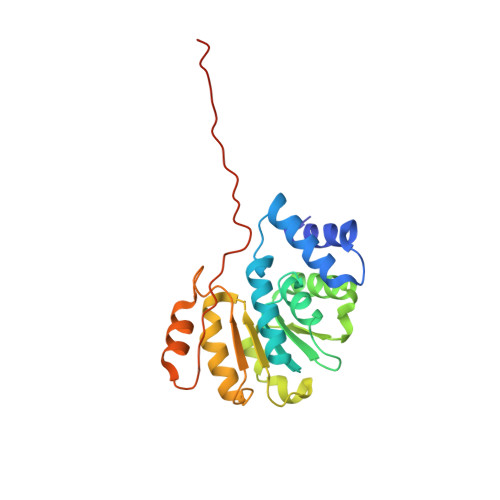
Rat catechol O-methyltransferase in complex with a catechol-type, pyridylsulfanyl-containing inhibitor - humanized form


Experimental Data Snapshot

  • Method: X-RAY DIFFRACTION
  • Resolution: 1.52 Å
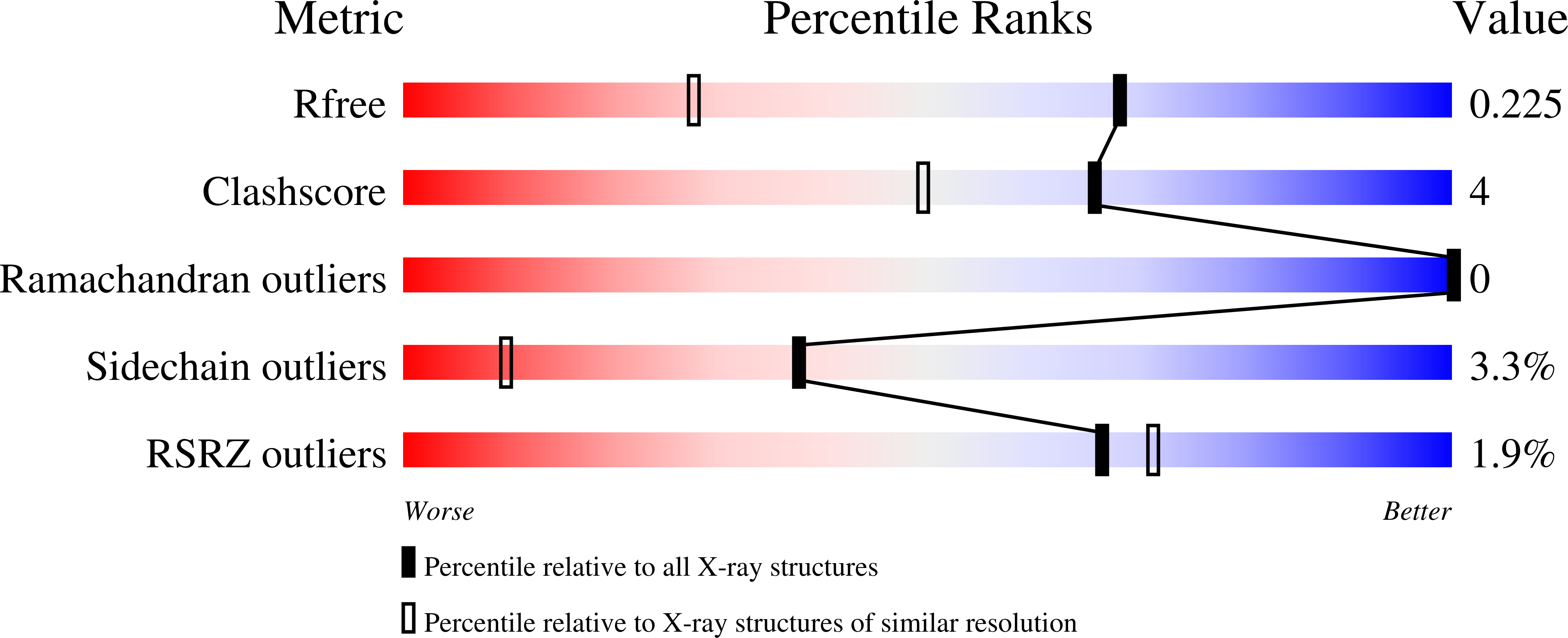
  • R-Value Free: 0.221 
  • R-Value Work: 0.193 
  • R-Value Observed: 0.194 

Starting Model: other
View more details

wwPDB Validation   3D Report Full Report


Ligand Structure Quality Assessment 


This is version 1.6 of the entry. See complete history


Literature

Molecular Recognition at the Active Site of Catechol-O-methyltransferase (COMT): Adenine Replacements in Bisubstrate Inhibitors

Ellermann, M.Paulini, R.Jakob-Roetne, R.Lerner, C.Borroni, E.Roth, D.Ehler, A.Schweizer, W.B.Schlatter, D.Rudolph, M.G.Diederich, F.

(2011) Chemistry 17: 6369-6381

  • DOI: https://doi.org/10.1002/chem.201003648
  • Primary Citation of Related Structures:  
    3NW9, 3OE4, 3OE5, 3OZR, 3OZS, 3OZT

  • PubMed Abstract: 

    L-Dopa, the standard therapeutic for Parkinson's disease, is inactivated by the enzyme catechol-O-methyltransferase (COMT). COMT catalyzes the transfer of an activated methyl group from S-adenosylmethionine (SAM) to its catechol substrates, such as L-dopa, in the presence of magnesium ions. The molecular recognition properties of the SAM-binding site of COMT have been investigated only sparsely. Here, we explore this site by structural alterations of the adenine moiety of bisubstrate inhibitors. The molecular recognition of adenine is of special interest due to the great abundance and importance of this nucleobase in biological systems. Novel bisubstrate inhibitors with adenine replacements were developed by structure-based design and synthesized using a nucleosidation protocol introduced by Vorbrüggen and co-workers. Key interactions of the adenine moiety with COMT were measured with a radiochemical assay. Several bisubstrate inhibitors, most notably the adenine replacements thiopyridine, purine, N-methyladenine, and 6-methylpurine, displayed nanomolar IC(50) values (median inhibitory concentration) for COMT down to 6 nM. A series of six cocrystal structures of the bisubstrate inhibitors in ternary complexes with COMT and Mg(2+) confirm our predicted binding mode of the adenine replacements. The cocrystal structure of an inhibitor bearing no nucleobase can be regarded as an intermediate along the reaction coordinate of bisubstrate inhibitor binding to COMT. Our studies show that solvation varies with the type of adenine replacement, whereas among the adenine derivatives, the nitrogen atom at position 1 is essential for high affinity, while the exocyclic amino group is most efficiently substituted by a methyl group.


  • Organizational Affiliation

    Laboratorium für Organische Chemie, ETH-Zürich, Wolfgang-Pauli-Strasse 10, 8093 Zürich, Switzerland.


Macromolecules
Find similar proteins by:  (by identity cutoff)  |  3D Structure
Entity ID: 1
MoleculeChains Sequence LengthOrganismDetailsImage
Catechol O-methyltransferase221Rattus norvegicusMutation(s): 2 
Gene Names: Comt
EC: 2.1.1.6
UniProt
Find proteins for P22734 (Rattus norvegicus)
Explore P22734 
Go to UniProtKB:  P22734
Entity Groups  
Sequence Clusters30% Identity50% Identity70% Identity90% Identity95% Identity100% Identity
UniProt GroupP22734
Sequence Annotations
Expand
  • Reference Sequence
Small Molecules
Ligands 2 Unique
IDChains Name / Formula / InChI Key2D Diagram3D Interactions
611
Query on 611

Download Ideal Coordinates CCD File 
C [auth A]N-[(E)-3-[(2R,3S,4R,5S)-3,4-dihydroxy-5-pyridin-4-ylsulfanyl-oxolan-2-yl]prop-2-enyl]-2,3-dihydroxy-5-nitro-benzamide
C19 H19 N3 O8 S
XRZIENNFYKGKSZ-GXSCOFJOSA-N
MG
Query on MG

Download Ideal Coordinates CCD File 
B [auth A]MAGNESIUM ION
Mg
JLVVSXFLKOJNIY-UHFFFAOYSA-N
Binding Affinity Annotations 
IDSourceBinding Affinity
611 PDBBind:  3OE5 Ki: 132 (nM) from 1 assay(s)
Experimental Data & Validation

Experimental Data

  • Method: X-RAY DIFFRACTION
  • Resolution: 1.52 Å
  • R-Value Free: 0.221 
  • R-Value Work: 0.193 
  • R-Value Observed: 0.194 
  • Space Group: P 32 2 1
Unit Cell:
Length ( Å )Angle ( ˚ )
a = 50.687α = 90
b = 50.687β = 90
c = 167.765γ = 120
Software Package:
Software NamePurpose
PHASERphasing
REFMACrefinement
XDSdata reduction
SADABSdata scaling

Structure Validation

View Full Validation Report


Ligand Structure Quality Assessment 


Entry History 

Deposition Data

Revision History  (Full details and data files)

  • Version 1.0: 2011-03-16
    Type: Initial release
  • Version 1.1: 2011-07-13
    Changes: Version format compliance
  • Version 1.2: 2012-05-23
    Changes: Structure summary
  • Version 1.3: 2012-08-15
    Changes: Structure summary
  • Version 1.4: 2017-11-08
    Changes: Refinement description
  • Version 1.5: 2024-03-20
    Changes: Data collection, Database references, Derived calculations
  • Version 1.6: 2024-04-03
    Changes: Refinement description