3ODT | pdb_00003odt

Crystal structure of WD40 beta propeller domain of Doa1


Experimental Data Snapshot

  • Method: X-RAY DIFFRACTION
  • Resolution: 1.35 Å
  • R-Value Free: 
    0.176 (Depositor), 0.199 (DCC) 
  • R-Value Work: 
    0.136 (Depositor) 
  • R-Value Observed: 
    0.139 (Depositor) 

wwPDB Validation 3D Report Full Report

Validation slider image for 3ODT

This is version 1.3 of the entry. See complete history

Literature

WD40 Repeat Propellers Define a Ubiquitin-Binding Domain that Regulates Turnover of F Box Proteins.

Pashkova, N.Gakhar, L.Winistorfer, S.C.Yu, L.Ramaswamy, S.Piper, R.C.

(2010) Mol Cell 40: 433-443

  • DOI: https://doi.org/10.1016/j.molcel.2010.10.018
  • Primary Citation Related Structures: 
    3ODT

  • PubMed Abstract: 

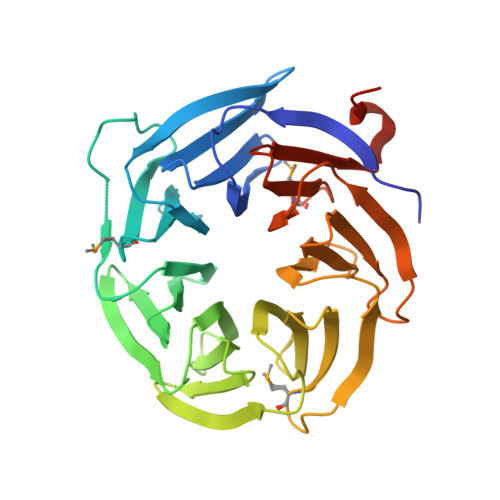
    WD40-repeat β-propellers are found in a wide range of proteins involved in distinct biological activities. We define a large subset of WD40 β-propellers as a class of ubiquitin-binding domains. Using the β-propeller from Doa1/Ufd3 as a paradigm, we find the conserved top surface of the Doa1 β-propeller binds the hydrophobic patch of ubiquitin centered on residues I44, L8, and V70. Mutations that disrupt ubiquitin binding abrogate Doa1 function, demonstrating the importance of this interaction. We further demonstrate that WD40 β-propellers from a functionally diverse set of proteins bind ubiquitin in a similar fashion. This set includes members of the F box family of SCF ubiquitin E3 ligase adaptors. Using mutants defective in binding, we find that ubiquitin interaction by the F box protein Cdc4 promotes its autoubiquitination and turnover. Collectively, our results reveal a molecular mechanism that may account for how ubiquitin controls a broad spectrum of cellular activities.


  • Organizational Affiliation
    • Department of Molecular Physiology and Biophysics, University of Iowa, Iowa City, IA 52242, USA.

Macromolecule Content 

  • Total Structure Weight: 69.58 kDa 
  • Atom Count: 5,704 
  • Modeled Residue Count: 592 
  • Deposited Residue Count: 626 
  • Unique protein chains: 1

Macromolecules

Find similar proteins by:|  3D Structure
Entity ID: 1
MoleculeChains  Sequence LengthOrganismDetailsImage
Protein DOA1
A, B
313Saccharomyces cerevisiaeMutation(s): 0 
Gene Names: DOA1UFD3ZZZ4YKL213C
UniProt
Find proteins for P36037 (Saccharomyces cerevisiae (strain ATCC 204508 / S288c))
Explore P36037 
Go to UniProtKB:  P36037
Entity Groups
Sequence Clusters30% Identity50% Identity70% Identity90% Identity95% Identity100% Identity
UniProt GroupP36037
Sequence Annotations
Expand
Reference Sequence

Experimental Data & Validation

Experimental Data

  • Method: X-RAY DIFFRACTION
  • Resolution: 1.35 Å
  • R-Value Free:  0.176 (Depositor), 0.199 (DCC) 
  • R-Value Work:  0.136 (Depositor) 
  • R-Value Observed: 0.139 (Depositor) 
Space Group: P 1 21 1
Unit Cell:
Length ( Å )Angle ( ˚ )
a = 33.656α = 90
b = 87.996β = 96.73
c = 92.265γ = 90
Software Package:
Software NamePurpose
Blu-Icedata collection
SOLVEphasing
PHENIXrefinement
d*TREKdata reduction
d*TREKdata scaling

Structure Validation

View Full Validation Report


Entry History 

Deposition Data

Revision History  (Full details and data files)

  • Version 1.0: 2010-12-01
    Type: Initial release
  • Version 1.1: 2011-07-13
    Changes: Version format compliance
  • Version 1.2: 2019-12-25
    Changes: Advisory, Database references, Derived calculations
  • Version 1.3: 2024-10-16
    Changes: Data collection, Database references, Derived calculations, Structure summary