3ODN | pdb_00003odn

The crystal structure of Drosophila Dally-Like Protein core domain


Experimental Data Snapshot

  • Method: X-RAY DIFFRACTION
  • Resolution: 2.40 Å
  • R-Value Free: 
    0.298 (Depositor), 0.309 (DCC) 
  • R-Value Work: 
    0.247 (Depositor), 0.257 (DCC) 
  • R-Value Observed: 
    0.249 (Depositor) 

wwPDB Validation 3D Report Full Report

Validation slider image for 3ODN

This is version 1.4 of the entry. See complete history

Literature

Structure of the protein core of the glypican Dally-like and localization of a region important for hedgehog signaling.

Kim, M.S.Saunders, A.M.Hamaoka, B.Y.Beachy, P.A.Leahy, D.J.

(2011) Proc Natl Acad Sci U S A 108: 13112-13117

  • DOI: https://doi.org/10.1073/pnas.1109877108
  • Primary Citation Related Structures: 
    3ODN

  • PubMed Abstract: 

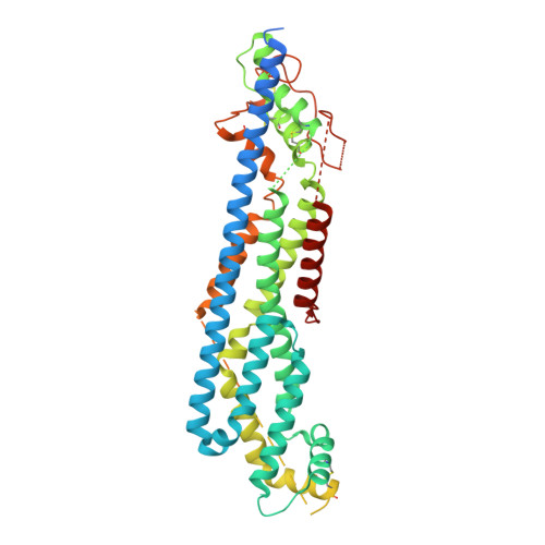
    Glypicans are heparan sulfate proteoglycans that modulate the signaling of multiple growth factors active during animal development, and loss of glypican function is associated with widespread developmental abnormalities. Glypicans consist of a conserved, approximately 45-kDa N-terminal protein core region followed by a stalk region that is tethered to the cell membrane by a glycosyl-phosphatidylinositol anchor. The stalk regions are predicted to be random coil but contain a variable number of attachment sites for heparan sulfate chains. Both the N-terminal protein core and the heparan sulfate attachments are important for glypican function. We report here the 2.4-Å crystal structure of the N-terminal protein core region of the Drosophila glypican Dally-like (Dlp). This structure reveals an elongated, α-helical fold for glypican core regions that does not appear homologous to any known structure. The Dlp core protein is required for normal responsiveness to Hedgehog (Hh) signals, and we identify a localized region on the Dlp surface important for mediating its function in Hh signaling. Purified Dlp protein core does not, however, interact appreciably with either Hh or an Hh:Ihog complex.


  • Organizational Affiliation
    • Department of Biophysics and Biophysical Chemistry, Johns Hopkins University School of Medicine, Baltimore, MD 21205, USA.

Macromolecule Content 

  • Total Structure Weight: 57.65 kDa 
  • Atom Count: 3,003 
  • Modeled Residue Count: 366 
  • Deposited Residue Count: 506 
  • Unique protein chains: 1

Macromolecules

Find similar proteins by:|  3D Structure
Entity ID: 1
MoleculeChains  Sequence LengthOrganismDetailsImage
Dally-like protein506Drosophila melanogasterMutation(s): 2 
Gene Names: dlpCG32146
UniProt
Find proteins for Q9VUG1 (Drosophila melanogaster)
Explore Q9VUG1 
Go to UniProtKB:  Q9VUG1
Entity Groups
Sequence Clusters30% Identity50% Identity70% Identity90% Identity95% Identity100% Identity
UniProt GroupQ9VUG1
Sequence Annotations
Expand
Reference Sequence

Experimental Data & Validation

Experimental Data

  • Method: X-RAY DIFFRACTION
  • Resolution: 2.40 Å
  • R-Value Free:  0.298 (Depositor), 0.309 (DCC) 
  • R-Value Work:  0.247 (Depositor), 0.257 (DCC) 
  • R-Value Observed: 0.249 (Depositor) 
Space Group: C 1 2 1
Unit Cell:
Length ( Å )Angle ( ˚ )
a = 97.021α = 90
b = 66.424β = 104.85
c = 85.725γ = 90
Software Package:
Software NamePurpose
HKL-2000data collection
SOLVEphasing
BUSTERrefinement
HKL-2000data reduction
HKL-2000data scaling

Structure Validation

View Full Validation Report


Entry History 

Deposition Data

Revision History  (Full details and data files)

  • Version 1.0: 2011-07-27
    Type: Initial release
  • Version 1.1: 2011-08-24
    Changes: Database references
  • Version 1.2: 2011-12-21
    Changes: Database references
  • Version 1.3: 2017-08-23
    Changes: Refinement description, Source and taxonomy
  • Version 1.4: 2024-10-30
    Changes: Data collection, Database references, Structure summary