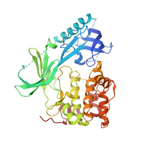
3O96

Crystal Structure of Human AKT1 with an Allosteric Inhibitor


Experimental Data Snapshot

  • Method: X-RAY DIFFRACTION
  • Resolution: 2.70 Å
  • R-Value Free: 0.308 
  • R-Value Work: 0.245 
  • R-Value Observed: 0.249 

Starting Models: experimental
View more details

wwPDB Validation   3D Report Full Report


Ligand Structure Quality Assessment 


This is version 1.3 of the entry. See complete history


Literature

Crystal structure of human AKT1 with an allosteric inhibitor reveals a new mode of kinase inhibition.

Wu, W.I.Voegtli, W.C.Sturgis, H.L.Dizon, F.P.Vigers, G.P.Brandhuber, B.J.

(2010) PLoS One 5: 12913-12913

  • DOI: https://doi.org/10.1371/journal.pone.0012913
  • Primary Citation of Related Structures:  
    3O96

  • PubMed Abstract: 

    AKT1 (NP_005154.2) is a member of the serine/threonine AGC protein kinase family involved in cellular metabolism, growth, proliferation and survival. The three human AKT isozymes are highly homologous multi-domain proteins with both overlapping and distinct cellular functions. Dysregulation of the AKT pathway has been identified in multiple human cancers. Several clinical trials are in progress to test the efficacy of AKT pathway inhibitors in treating cancer. Recently, a series of AKT isozyme-selective allosteric inhibitors have been reported. They require the presence of both the pleckstrin-homology (PH) and kinase domains of AKT, but their binding mode has not yet been elucidated. We present here a 2.7 Å resolution co-crystal structure of human AKT1 containing both the PH and kinase domains with a selective allosteric inhibitor bound in the interface. The structure reveals the interactions between the PH and kinase domains, as well as the critical amino residues that mediate binding of the inhibitor to AKT1. Our work also reveals an intricate balance in the enzymatic regulation of AKT, where the PH domain appears to lock the kinase in an inactive conformation and the kinase domain disrupts the phospholipid binding site of the PH domain. This information advances our knowledge in AKT1 structure and regulation, thereby providing a structural foundation for interpreting the effects of different classes of AKT inhibitors and designing selective ones.


  • Organizational Affiliation

    Department of Structural Biology, Array BioPharma Inc., Boulder, Colorado, United States of America.


Macromolecules
Find similar proteins by:  (by identity cutoff)  |  3D Structure
Entity ID: 1
MoleculeChains Sequence LengthOrganismDetailsImage
RAC-alpha serine/threonine-protein kinase446Homo sapiensMutation(s): 0 
Gene Names: AKT1PKBRAC
EC: 2.7.11.1
UniProt & NIH Common Fund Data Resources
Find proteins for P31749 (Homo sapiens)
Explore P31749 
Go to UniProtKB:  P31749
PHAROS:  P31749
GTEx:  ENSG00000142208 
Entity Groups  
Sequence Clusters30% Identity50% Identity70% Identity90% Identity95% Identity100% Identity
UniProt GroupP31749
Sequence Annotations
Expand
  • Reference Sequence
Small Molecules
Ligands 1 Unique
IDChains Name / Formula / InChI Key2D Diagram3D Interactions
IQO
Query on IQO

Download Ideal Coordinates CCD File 
B [auth A]1-(1-(4-(7-phenyl-1H-imidazo[4,5-g]quinoxalin-6-yl)benzyl)piperidin-4-yl)-1H-benzo[d]imidazol-2(3H)-one
C34 H29 N7 O
BIWGYFZAEWGBAL-UHFFFAOYSA-N
Binding Affinity Annotations 
IDSourceBinding Affinity
IQO BindingDB:  3O96 IC50: min: 58, max: 305 (nM) from 3 assay(s)
PDBBind:  3O96 IC50: 58 (nM) from 1 assay(s)
Experimental Data & Validation

Experimental Data

  • Method: X-RAY DIFFRACTION
  • Resolution: 2.70 Å
  • R-Value Free: 0.308 
  • R-Value Work: 0.245 
  • R-Value Observed: 0.249 
  • Space Group: P 1 21 1
Unit Cell:
Length ( Å )Angle ( ˚ )
a = 49.31α = 90
b = 69.94β = 100.6
c = 61.85γ = 90
Software Package:
Software NamePurpose
CrystalCleardata collection
MOLREPphasing
CNXrefinement
MOSFLMdata reduction
SCALAdata scaling

Structure Validation

View Full Validation Report


Ligand Structure Quality Assessment 


Entry History 

Deposition Data

Revision History  (Full details and data files)

  • Version 1.0: 2010-10-13
    Type: Initial release
  • Version 1.1: 2011-07-13
    Changes: Version format compliance
  • Version 1.2: 2023-09-06
    Changes: Data collection, Database references, Derived calculations, Refinement description
  • Version 1.3: 2024-10-16
    Changes: Structure summary