3O8J | pdb_00003o8j

Crystal structure of 2-methylcitrate synthase (PrpC) from Salmonella typhimurium


Experimental Data Snapshot

  • Method: X-RAY DIFFRACTION
  • Resolution: 2.41 Å
  • R-Value Free: 
    0.282 (Depositor), 0.282 (DCC) 
  • R-Value Work: 
    0.218 (Depositor), 0.220 (DCC) 
  • R-Value Observed: 
    0.221 (Depositor) 

Starting Model: experimental
View more details

wwPDB Validation 3D Report Full Report

Validation slider image for 3O8J

This is version 1.2 of the entry. See complete history

Literature

Crystal structure of Salmonella typhimurium 2-methylcitrate synthase: Insights on domain movement and substrate specificity

Chittori, S.Savithri, H.S.Murthy, M.R.N.

(2011) J Struct Biol 174: 58-68

  • DOI: https://doi.org/10.1016/j.jsb.2010.10.008
  • Primary Citation Related Structures: 
    3O8J

  • PubMed Abstract: 

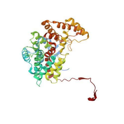
    2-Methylcitric acid (2-MCA) cycle is one of the well studied pathways for the utilization of propionate as a source of carbon and energy in bacteria such as Salmonella typhimurium and Escherichia coli. 2-Methylcitrate synthase (2-MCS) catalyzes the conversion of oxaloacetate and propionyl-CoA to 2-methylcitrate and CoA in the second step of 2-MCA cycle. Here, we report the X-ray crystal structure of S. typhimurium 2-MCS (StPrpC) at 2.4Å resolution and its functional characterization. StPrpC was found to utilize propionyl-CoA more efficiently than acetyl-CoA or butyryl-CoA. The polypeptide fold and the catalytic residues of StPrpC are conserved in citrate synthases (CSs) suggesting similarities in their functional mechanisms. In the triclinic P1 cell, StPrpC molecules were organized as decamers composed of five identical dimer units. In solution, StPrpC was in a dimeric form at low concentrations and was converted to larger oligomers at higher concentrations. CSs are usually dimeric proteins. In Gram-negative bacteria, a hexameric form, believed to be important for regulation of activity by NADH, is also observed. Structural comparisons with hexameric E. coli CS suggested that the key residues involved in NADH binding are not conserved in StPrpC. Structural comparison with the ligand free and bound states of CSs showed that StPrpC is in a nearly closed conformation despite the absence of bound ligands. It was found that the Tyr197 and Leu324 of StPrpC are structurally equivalent to the ligand binding residues His and Val, respectively, of CSs. These substitutions might determine the specificities for acyl-CoAs of these enzymes.


  • Organizational Affiliation
    • Molecular Biophysics Unit, Indian Institute of Science, Bangalore, Karnataka 560012, India.

Macromolecule Content 

  • Total Structure Weight: 450.8 kDa 
  • Atom Count: 29,229 
  • Modeled Residue Count: 3,624 
  • Deposited Residue Count: 4,040 
  • Unique protein chains: 1

Macromolecules

Find similar proteins by:|  3D Structure
Entity ID: 1
MoleculeChains  Sequence LengthOrganismDetailsImage
2-methylcitrate synthase
A, B, C, D, E
A, B, C, D, E, F, G, H, I, J
404Salmonella enterica subsp. enterica serovar TyphimuriumMutation(s): 0 
Gene Names: prpCSTM0369
EC: 2.3.3.5 (PDB Primary Data), 2.3.3.16 (UniProt)
UniProt
Find proteins for Q56063 (Salmonella typhimurium (strain LT2 / SGSC1412 / ATCC 700720))
Explore Q56063 
Go to UniProtKB:  Q56063
Entity Groups
Sequence Clusters30% Identity50% Identity70% Identity90% Identity95% Identity100% Identity
UniProt GroupQ56063
Sequence Annotations
Expand
Reference Sequence

Small Molecules

Ligands 1 Unique
IDChains Name / Formula / InChI Key2D Diagram3D Interactions
GOL

Query on GOL


Download:Ideal Coordinates CCD File
K [auth A]
L [auth B]
M [auth C]
N [auth E]
O [auth E]
K [auth A],
L [auth B],
M [auth C],
N [auth E],
O [auth E],
P [auth F],
Q [auth F],
R [auth G],
S [auth H],
T [auth I],
U [auth J]
GLYCEROL
C3 H8 O3
PEDCQBHIVMGVHV-UHFFFAOYSA-N

Experimental Data & Validation

Experimental Data

  • Method: X-RAY DIFFRACTION
  • Resolution: 2.41 Å
  • R-Value Free:  0.282 (Depositor), 0.282 (DCC) 
  • R-Value Work:  0.218 (Depositor), 0.220 (DCC) 
  • R-Value Observed: 0.221 (Depositor) 
Space Group: P 1
Unit Cell:
Length ( Å )Angle ( ˚ )
a = 92.068α = 60.84
b = 118.159β = 67.77
c = 120.659γ = 81.92
Software Package:
Software NamePurpose
MAR345dtbdata collection
PHASERphasing
REFMACrefinement
HKL-2000data reduction
SCALEPACKdata scaling

Structure Validation

View Full Validation Report


Entry History 

Deposition Data

Revision History  (Full details and data files)

  • Version 1.0: 2011-04-13
    Type: Initial release
  • Version 1.1: 2011-07-13
    Changes: Version format compliance
  • Version 1.2: 2023-11-01
    Changes: Data collection, Database references, Derived calculations, Refinement description