3O7L | pdb_00003o7l

Crystal Structure of phospholamban (1-19):PKA C-subunit:AMP-PNP:Mg2+ complex


Experimental Data Snapshot

  • Method: X-RAY DIFFRACTION
  • Resolution: 2.80 Å
  • R-Value Free: 
    0.287 (Depositor) 
  • R-Value Work: 
    0.216 (Depositor), 0.231 (DCC) 
  • R-Value Observed: 
    0.216 (Depositor) 

Starting Model: experimental
View more details

wwPDB Validation 3D Report Full Report

Validation slider image for 3O7L

Ligand Structure Quality Assessment 


This is version 1.3 of the entry. See complete history

Literature

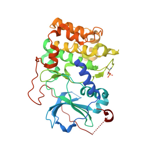
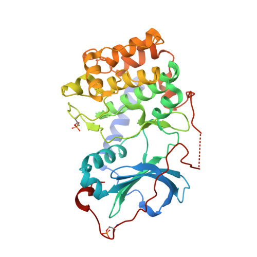
Dynamics connect substrate recognition to catalysis in protein kinase A.

Masterson, L.R.Cheng, C.Yu, T.Tonelli, M.Kornev, A.Taylor, S.S.Veglia, G.

(2010) Nat Chem Biol 6: 821-828

  • DOI: https://doi.org/10.1038/nchembio.452
  • Primary Citation Related Structures: 
    3O7L

  • PubMed Abstract: 

    Atomic resolution studies of protein kinases have traditionally been carried out in the inhibitory state, limiting our current knowledge on the mechanisms of substrate recognition and catalysis. Using NMR, X-ray crystallography and thermodynamic measurements, we analyzed the substrate recognition process of cAMP-dependent protein kinase (PKA), finding that entropy and protein dynamics play a prominent role. The nucleotide acts as a dynamic and allosteric activator by coupling the two lobes of apo PKA, enhancing the enzyme dynamics synchronously and priming it for catalysis. The formation of the ternary complex is entropically driven, and NMR spin relaxation data reveal that both substrate and PKA are dynamic in the closed state. Our results show that the enzyme toggles between open and closed states, which indicates that a conformational selection rather than an induced-fit mechanism governs substrate recognition.


  • Organizational Affiliation
    • Department of Biochemistry, Molecular Biology and Biophysics, University of Minnesota, Minneapolis, Minnesota, USA.

Macromolecule Content 

  • Total Structure Weight: 83.63 kDa 
  • Atom Count: 5,231 
  • Modeled Residue Count: 677 
  • Deposited Residue Count: 715 
  • Unique protein chains: 3

Macromolecules

Find similar proteins by:|  3D Structure
Entity ID: 1
MoleculeChains  Sequence LengthOrganismDetailsImage
cAMP-dependent protein kinase catalytic subunit alphaA [auth B]350Mus musculusMutation(s): 0 
Gene Names: PkacaPrkaca
EC: 2.7.11.11
UniProt
Find proteins for P05132 (Mus musculus)
Explore P05132 
Go to UniProtKB:  P05132
Entity Groups
Sequence Clusters30% Identity50% Identity70% Identity90% Identity95% Identity100% Identity
UniProt GroupP05132
Sequence Annotations
Expand
Reference Sequence
Find similar proteins by:|  3D Structure
Entity ID: 2
MoleculeChains  Sequence LengthOrganismDetailsImage
cAMP-dependent protein kinase catalytic subunit alphaB [auth D]350Mus musculusMutation(s): 0 
Gene Names: PkacaPrkaca
EC: 2.7.11.11
UniProt
Find proteins for P05132 (Mus musculus)
Explore P05132 
Go to UniProtKB:  P05132
Entity Groups
Sequence Clusters30% Identity50% Identity70% Identity90% Identity95% Identity100% Identity
UniProt GroupP05132
Sequence Annotations
Expand
Reference Sequence
Find similar proteins by:  Sequence   |   3D Structure  
Entity ID: 3
MoleculeChains  Sequence LengthOrganismDetailsImage
Cardiac phospholambanC [auth I]15N/AMutation(s): 0 
UniProt
Find proteins for P61014 (Mus musculus)
Explore P61014 
Go to UniProtKB:  P61014
Entity Groups
Sequence Clusters30% Identity50% Identity70% Identity90% Identity95% Identity100% Identity
UniProt GroupP61014
Sequence Annotations
Expand
Reference Sequence

Small Molecules

Modified Residues  2 Unique
IDChains TypeFormula2D DiagramParent
TPO
Query on TPO
A [auth B]L-PEPTIDE LINKINGC4 H10 N O6 PTHR
SEP
Query on SEP
B [auth D]L-PEPTIDE LINKINGC3 H8 N O6 PSER

Experimental Data & Validation

Experimental Data

  • Method: X-RAY DIFFRACTION
  • Resolution: 2.80 Å
  • R-Value Free:  0.287 (Depositor) 
  • R-Value Work:  0.216 (Depositor), 0.231 (DCC) 
  • R-Value Observed: 0.216 (Depositor) 
Space Group: P 61
Unit Cell:
Length ( Å )Angle ( ˚ )
a = 92.193α = 90
b = 92.193β = 90
c = 192.186γ = 120
Software Package:
Software NamePurpose
ADSCdata collection
PHASESphasing
PHENIXrefinement
HKL-2000data reduction
HKL-2000data scaling

Structure Validation

View Full Validation Report


Ligand Structure Quality Assessment 


Entry History 

Deposition Data

Revision History  (Full details and data files)

  • Version 1.0: 2010-10-06
    Type: Initial release
  • Version 1.1: 2011-07-13
    Changes: Version format compliance
  • Version 1.2: 2023-09-06
    Changes: Data collection, Database references, Derived calculations, Refinement description
  • Version 1.3: 2024-10-16
    Changes: Structure summary