3O49 | pdb_00003o49

Crystal structure of Symfoil-1: de novo designed beta-trefoil architecture with symmetric primary structure


Experimental Data Snapshot

  • Method: X-RAY DIFFRACTION
  • Resolution: 1.45 Å
  • R-Value Free: 
    0.227 (Depositor) 
  • R-Value Work: 
    0.196 (Depositor) 
  • R-Value Observed: 
    0.199 (Depositor) 

wwPDB Validation 3D Report Full Report

Validation slider image for 3O49

This is version 1.2 of the entry. See complete history

Literature

Experimental support for the evolution of symmetric protein architecture from a simple peptide motif.

Lee, J.Blaber, M.

(2011) Proc Natl Acad Sci U S A 108: 126-130

  • DOI: https://doi.org/10.1073/pnas.1015032108
  • Primary Citation Related Structures: 
    3O49, 3O4A, 3O4B, 3O4C, 3O4D, 3OGF, 3OL0

  • PubMed Abstract: 

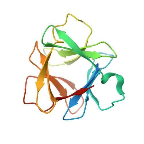
    The majority of protein architectures exhibit elements of structural symmetry, and "gene duplication and fusion" is the evolutionary mechanism generally hypothesized to be responsible for their emergence from simple peptide motifs. Despite the central importance of the gene duplication and fusion hypothesis, experimental support for a plausible evolutionary pathway for a specific protein architecture has yet to be effectively demonstrated. To address this question, a unique "top-down symmetric deconstruction" strategy was utilized to successfully identify a simple peptide motif capable of recapitulating, via gene duplication and fusion processes, a symmetric protein architecture (the threefold symmetric β-trefoil fold). The folding properties of intermediary forms in this deconstruction agree precisely with a previously proposed "conserved architecture" model for symmetric protein evolution. Furthermore, a route through foldable sequence-space between the simple peptide motif and extant protein fold is demonstrated. These results provide compelling experimental support for a plausible evolutionary pathway of symmetric protein architecture via gene duplication and fusion processes.


  • Organizational Affiliation
    • Department of Biomedical Sciences, Florida State University, Tallahassee FL 32306-4300, USA.

Macromolecule Content 

  • Total Structure Weight: 15.96 kDa 
  • Atom Count: 1,163 
  • Modeled Residue Count: 123 
  • Deposited Residue Count: 142 
  • Unique protein chains: 1

Macromolecules

Find similar proteins by:|  3D Structure
Entity ID: 1
MoleculeChains  Sequence LengthOrganismDetailsImage
de novo designed beta-trefoil architecture with symmetric primary structure142synthetic constructMutation(s): 0 

Small Molecules

Ligands 2 Unique
IDChains Name / Formula / InChI Key2D Diagram3D Interactions
TRS

Query on TRS


Download:Ideal Coordinates CCD File
B [auth A]2-AMINO-2-HYDROXYMETHYL-PROPANE-1,3-DIOL
C4 H12 N O3
LENZDBCJOHFCAS-UHFFFAOYSA-O
SO4

Query on SO4


Download:Ideal Coordinates CCD File
C [auth A]SULFATE ION
O4 S
QAOWNCQODCNURD-UHFFFAOYSA-L

Experimental Data & Validation

Experimental Data

  • Method: X-RAY DIFFRACTION
  • Resolution: 1.45 Å
  • R-Value Free:  0.227 (Depositor) 
  • R-Value Work:  0.196 (Depositor) 
  • R-Value Observed: 0.199 (Depositor) 
Space Group: I 2 2 2
Unit Cell:
Length ( Å )Angle ( ˚ )
a = 50.365α = 90
b = 53.351β = 90
c = 85.175γ = 90
Software Package:
Software NamePurpose
HKL-2000data collection
PHASESphasing
PHENIXrefinement
HKL-2000data reduction
HKL-2000data scaling

Structure Validation

View Full Validation Report


Entry History 

Deposition Data

Revision History  (Full details and data files)

  • Version 1.0: 2010-12-22
    Type: Initial release
  • Version 1.1: 2011-07-13
    Changes: Version format compliance
  • Version 1.2: 2024-02-21
    Changes: Data collection, Database references, Derived calculations