3O3X | pdb_00003o3x

Crystal structure of gp41-5, a single-chain 5-helix-bundle based on HIV gp41


Experimental Data Snapshot

  • Method: X-RAY DIFFRACTION
  • Resolution: 1.45 Å
  • R-Value Free: 
    0.181 (Depositor), 0.182 (DCC) 
  • R-Value Work: 
    0.150 (Depositor), 0.151 (DCC) 
  • R-Value Observed: 
    0.152 (Depositor) 

Starting Model: experimental
View more details

wwPDB Validation 3D Report Full Report

Validation slider image for 3O3X

This is version 1.1 of the entry. See complete history

Literature

Broad Distribution of Energetically Important Contacts across an Extended Protein Interface.

Johnson, L.M.Horne, W.S.Gellman, S.H.

(2011) J Am Chem Soc 133: 10038-10041

  • DOI: https://doi.org/10.1021/ja203358t
  • Primary Citation Related Structures: 
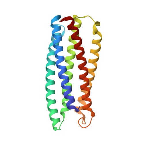
    3O3X, 3O3Z, 3O40, 3O43

  • PubMed Abstract: 

    Infection of cells by HIV depends upon profound structural rearrangements within the trimeric viral protein gp41. Critical to this process is the formation of a six-helix bundle in which a set of three N-terminal heptad repeat (NHR) helices assemble to form a core displaying long grooves that provide docking sites for three C-terminal heptad repeat (CHR) helices. We report experiments designed to discriminate between two alternative hypotheses regarding the source of affinity between individual CHR helices and the complementary groove: (1) affinity is dominated by interactions of a small cluster of side chains at one end of the CHR helix; or (2) affinity depends upon interactions distributed across the long CHR helix. We have employed two complementary experimental designs, and results from both favor the latter hypothesis.


  • Organizational Affiliation
    • Department of Chemistry, University of Wisconsin, Madison, Wisconsin 53706, USA.

Macromolecule Content 

  • Total Structure Weight: 22.43 kDa 
  • Atom Count: 1,883 
  • Modeled Residue Count: 198 
  • Deposited Residue Count: 198 
  • Unique protein chains: 1

Macromolecules

Find similar proteins by:|  3D Structure
Entity ID: 1
MoleculeChains  Sequence LengthOrganismDetailsImage
gp41-5198synthetic constructMutation(s): 0 

Small Molecules

Experimental Data & Validation

Experimental Data

  • Method: X-RAY DIFFRACTION
  • Resolution: 1.45 Å
  • R-Value Free:  0.181 (Depositor), 0.182 (DCC) 
  • R-Value Work:  0.150 (Depositor), 0.151 (DCC) 
  • R-Value Observed: 0.152 (Depositor) 
Space Group: P 21 21 21
Unit Cell:
Length ( Å )Angle ( ˚ )
a = 54.459α = 90
b = 57.936β = 90
c = 61.689γ = 90
Software Package:
Software NamePurpose
MD2data collection
PHASERphasing
REFMACrefinement
HKL-2000data reduction
HKL-2000data scaling

Structure Validation

View Full Validation Report


Entry History 

Deposition Data

Revision History  (Full details and data files)

  • Version 1.0: 2011-07-27
    Type: Initial release
  • Version 1.1: 2023-09-06
    Changes: Data collection, Database references, Derived calculations, Refinement description