3O2Q

Crystal structure of the human symplekin-Ssu72-CTD phosphopeptide complex


Experimental Data Snapshot

  • Method: X-RAY DIFFRACTION
  • Resolution: 2.40 Å
  • R-Value Free: 0.242 
  • R-Value Work: 0.184 
  • R-Value Observed: 0.187 

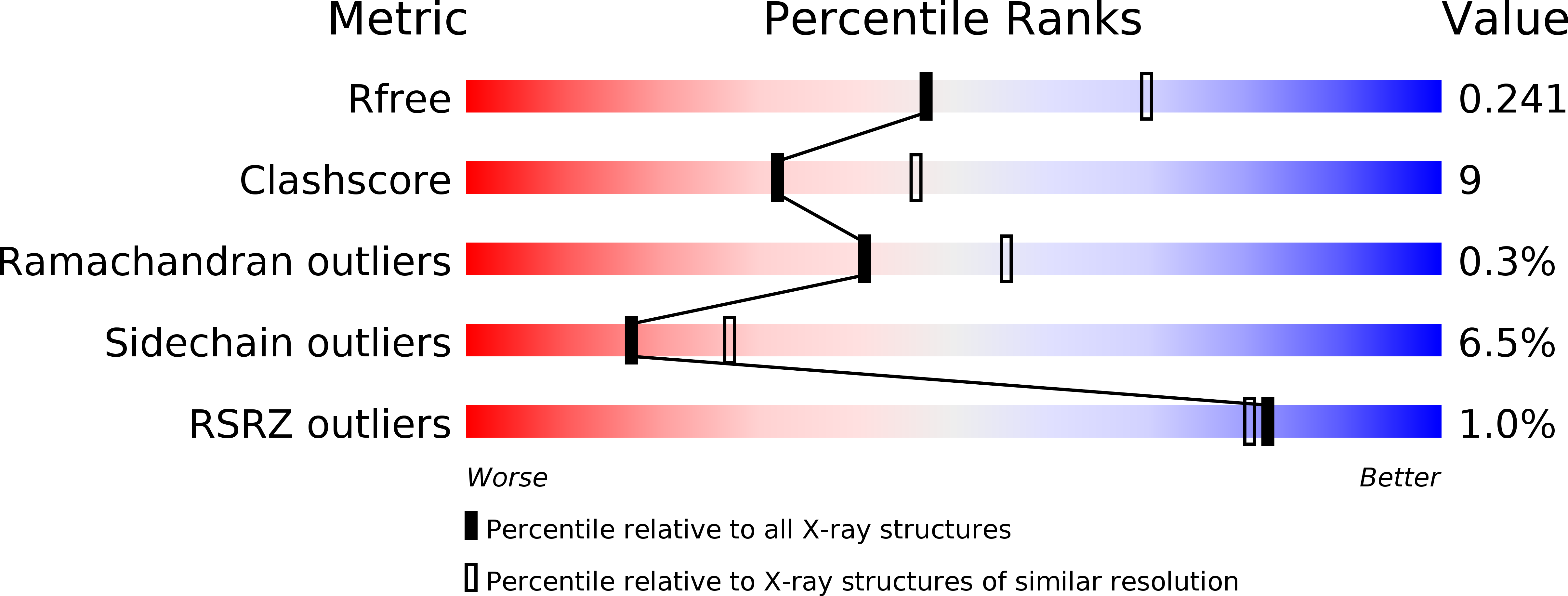
wwPDB Validation   3D Report Full Report


This is version 1.3 of the entry. See complete history


Literature

Crystal structure of the human symplekin-Ssu72-CTD phosphopeptide complex.

Xiang, K.Nagaike, T.Xiang, S.Kilic, T.Beh, M.M.Manley, J.L.Tong, L.

(2010) Nature 467: 729-733

  • DOI: https://doi.org/10.1038/nature09391
  • Primary Citation of Related Structures:  
    3O2Q, 3O2S, 3O2T, 3ODR, 3ODS

  • PubMed Abstract: 

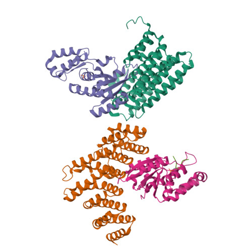
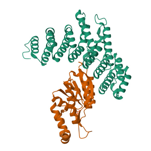
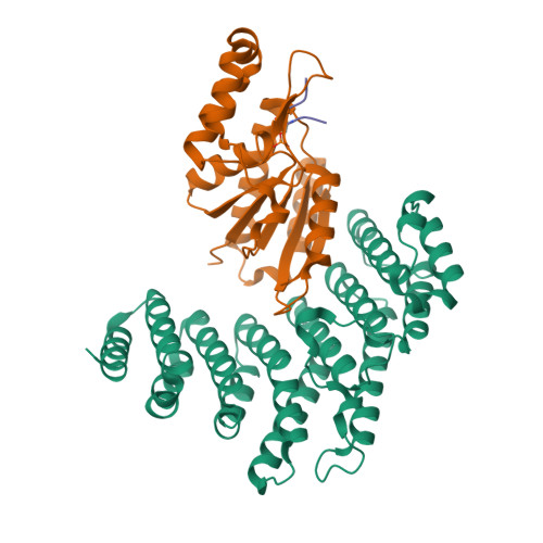
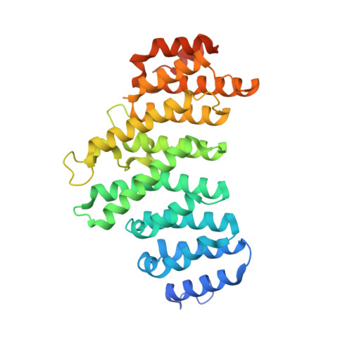
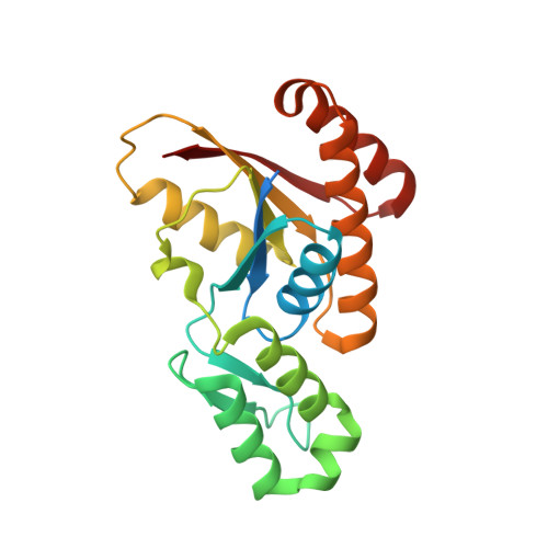
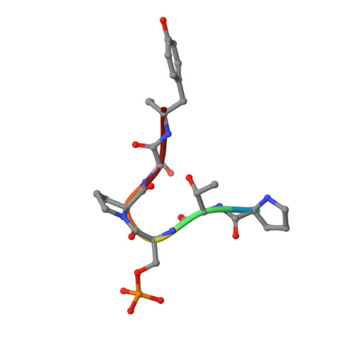
    Symplekin (Pta1 in yeast) is a scaffold in the large protein complex that is required for 3'-end cleavage and polyadenylation of eukaryotic messenger RNA precursors (pre-mRNAs); it also participates in transcription initiation and termination by RNA polymerase II (Pol II). Symplekin mediates interactions between many different proteins in this machinery, although the molecular basis for its function is not known. Here we report the crystal structure at 2.4 Å resolution of the amino-terminal domain (residues 30-340) of human symplekin in a ternary complex with the Pol II carboxy-terminal domain (CTD) Ser 5 phosphatase Ssu72 (refs 7, 10-17) and a CTD Ser 5 phosphopeptide. The N-terminal domain of symplekin has the ARM or HEAT fold, with seven pairs of antiparallel α-helices arranged in the shape of an arc. The structure of Ssu72 has some similarity to that of low-molecular-mass phosphotyrosine protein phosphatase, although Ssu72 has a unique active-site landscape as well as extra structural features at the C terminus that are important for interaction with symplekin. Ssu72 is bound to the concave face of symplekin, and engineered mutations in this interface can abolish interactions between the two proteins. The CTD peptide is bound in the active site of Ssu72, with the pSer 5-Pro 6 peptide bond in the cis configuration, which contrasts with all other known CTD peptide conformations. Although the active site of Ssu72 is about 25 Å from the interface with symplekin, we found that the symplekin N-terminal domain stimulates Ssu72 CTD phosphatase activity in vitro. Furthermore, the N-terminal domain of symplekin inhibits polyadenylation in vitro, but only when coupled to transcription. Because catalytically active Ssu72 overcomes this inhibition, our results show a role for mammalian Ssu72 in transcription-coupled pre-mRNA 3'-end processing.


  • Organizational Affiliation

    Department of Biological Sciences, Columbia University, New York, New York 10027, USA.


Macromolecules
Find similar proteins by:  (by identity cutoff)  |  3D Structure
Entity ID: 1
MoleculeChains Sequence LengthOrganismDetailsImage
SymplekinA,
C [auth D]
351Homo sapiensMutation(s): 0 
Gene Names: SYMPKSPK
UniProt & NIH Common Fund Data Resources
Find proteins for Q92797 (Homo sapiens)
Explore Q92797 
Go to UniProtKB:  Q92797
PHAROS:  Q92797
GTEx:  ENSG00000125755 
Entity Groups  
Sequence Clusters30% Identity50% Identity70% Identity90% Identity95% Identity100% Identity
UniProt GroupQ92797
Sequence Annotations
Expand
  • Reference Sequence
Find similar proteins by:  (by identity cutoff)  |  3D Structure
Entity ID: 2
MoleculeChains Sequence LengthOrganismDetailsImage
RNA polymerase II subunit A C-terminal domain phosphatase SSU72B,
D [auth E]
214Homo sapiensMutation(s): 1 
Gene Names: SSU72HSPC182PNAS-120
EC: 3.1.3.16
UniProt & NIH Common Fund Data Resources
Find proteins for Q9NP77 (Homo sapiens)
Explore Q9NP77 
Go to UniProtKB:  Q9NP77
PHAROS:  Q9NP77
GTEx:  ENSG00000160075 
Entity Groups  
Sequence Clusters30% Identity50% Identity70% Identity90% Identity95% Identity100% Identity
UniProt GroupQ9NP77
Sequence Annotations
Expand
  • Reference Sequence

Find similar proteins by:  Sequence   |   3D Structure  

Entity ID: 3
MoleculeChains Sequence LengthOrganismDetailsImage
RNA polymerase II CTD Serine-5 phosphopeptideE [auth F]6N/AMutation(s): 0 
Sequence Annotations
Expand
  • Reference Sequence
Small Molecules
Ligands 1 Unique
IDChains Name / Formula / InChI Key2D Diagram3D Interactions
PO4
Query on PO4

Download Ideal Coordinates CCD File 
F [auth B]PHOSPHATE ION
O4 P
NBIIXXVUZAFLBC-UHFFFAOYSA-K
Modified Residues  1 Unique
IDChains TypeFormula2D DiagramParent
SEP
Query on SEP
E [auth F]L-PEPTIDE LINKINGC3 H8 N O6 PSER
Experimental Data & Validation

Experimental Data

  • Method: X-RAY DIFFRACTION
  • Resolution: 2.40 Å
  • R-Value Free: 0.242 
  • R-Value Work: 0.184 
  • R-Value Observed: 0.187 
  • Space Group: P 1 21 1
Unit Cell:
Length ( Å )Angle ( ˚ )
a = 67.045α = 90
b = 97.581β = 98.66
c = 105.008γ = 90
Software Package:
Software NamePurpose
CBASSdata collection
REFMACrefinement
HKL-2000data reduction
HKL-2000data scaling

Structure Validation

View Full Validation Report


Entry History 

Deposition Data

Revision History  (Full details and data files)

  • Version 1.0: 2010-10-06
    Type: Initial release
  • Version 1.1: 2011-07-13
    Changes: Version format compliance
  • Version 1.2: 2017-11-08
    Changes: Refinement description
  • Version 1.3: 2024-10-09
    Changes: Data collection, Database references, Derived calculations, Structure summary