3NZ1 | pdb_00003nz1

Crystal Structure of Kemp Elimination Catalyst 1A53-2 Complexed with Transition State Analog 5-Nitro Benzotriazole


Experimental Data Snapshot

  • Method: X-RAY DIFFRACTION
  • Resolution: 1.56 Å
  • R-Value Free: 
    0.210 (Depositor), 0.218 (DCC) 
  • R-Value Work: 
    0.172 (Depositor), 0.185 (DCC) 
  • R-Value Observed: 
    0.174 (Depositor) 

wwPDB Validation 3D Report Full Report

Validation slider image for 3NZ1

Ligand Structure Quality Assessment 


This is version 1.3 of the entry. See complete history

Literature

Iterative approach to computational enzyme design.

Privett, H.K.Kiss, G.Lee, T.M.Blomberg, R.Chica, R.A.Thomas, L.M.Hilvert, D.Houk, K.N.Mayo, S.L.

(2012) Proc Natl Acad Sci U S A 109: 3790-3795

  • DOI: https://doi.org/10.1073/pnas.1118082108
  • Primary Citation Related Structures: 
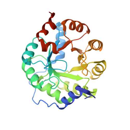
    3NYD, 3NYZ, 3NZ1, 3O2L

  • PubMed Abstract: 

    A general approach for the computational design of enzymes to catalyze arbitrary reactions is a goal at the forefront of the field of protein design. Recently, computationally designed enzymes have been produced for three chemical reactions through the synthesis and screening of a large number of variants. Here, we present an iterative approach that has led to the development of the most catalytically efficient computationally designed enzyme for the Kemp elimination to date. Previously established computational techniques were used to generate an initial design, HG-1, which was catalytically inactive. Analysis of HG-1 with molecular dynamics simulations (MD) and X-ray crystallography indicated that the inactivity might be due to bound waters and high flexibility of residues within the active site. This analysis guided changes to our design procedure, moved the design deeper into the interior of the protein, and resulted in an active Kemp eliminase, HG-2. The cocrystal structure of this enzyme with a transition state analog (TSA) revealed that the TSA was bound in the active site, interacted with the intended catalytic base in a catalytically relevant manner, but was flipped relative to the design model. MD analysis of HG-2 led to an additional point mutation, HG-3, that produced a further threefold improvement in activity. This iterative approach to computational enzyme design, including detailed MD and structural analysis of both active and inactive designs, promises a more complete understanding of the underlying principles of enzymatic catalysis and furthers progress toward reliably producing active enzymes.


  • Organizational Affiliation
    • Division of Chemistry and Chemical Engineering, California Institute of Technology, Pasadena, CA 91125, USA.

Macromolecule Content 

  • Total Structure Weight: 31.2 kDa 
  • Atom Count: 2,347 
  • Modeled Residue Count: 249 
  • Deposited Residue Count: 261 
  • Unique protein chains: 1

Macromolecules

Find similar proteins by:|  3D Structure
Entity ID: 1
MoleculeChains  Sequence LengthOrganismDetailsImage
Indole-3-glycerol phosphate synthase261Saccharolobus solfataricusMutation(s): 12 
Gene Names: INDOLE-3-GLYCEROLPHOSPHATE SYNTHASESSO0895trpC
EC: 4.1.1.48
UniProt
Find proteins for Q06121 (Saccharolobus solfataricus (strain ATCC 35092 / DSM 1617 / JCM 11322 / P2))
Explore Q06121 
Go to UniProtKB:  Q06121
Entity Groups
Sequence Clusters30% Identity50% Identity70% Identity90% Identity95% Identity100% Identity
UniProt GroupQ06121
Sequence Annotations
Expand
Reference Sequence

Small Molecules

Ligands 3 Unique
IDChains Name / Formula / InChI Key2D Diagram3D Interactions
3NY

Query on 3NY


Download:Ideal Coordinates CCD File
B [auth A]5-nitro-1H-benzotriazole
C6 H4 N4 O2
AOCDQWRMYHJTMY-UHFFFAOYSA-N
TLA

Query on TLA


Download:Ideal Coordinates CCD File
I [auth A],
J [auth A],
K [auth A]
L(+)-TARTARIC ACID
C4 H6 O6
FEWJPZIEWOKRBE-JCYAYHJZSA-N
SO4

Query on SO4


Download:Ideal Coordinates CCD File
C [auth A]
D [auth A]
E [auth A]
F [auth A]
G [auth A]
C [auth A],
D [auth A],
E [auth A],
F [auth A],
G [auth A],
H [auth A]
SULFATE ION
O4 S
QAOWNCQODCNURD-UHFFFAOYSA-L

Experimental Data & Validation

Experimental Data

  • Method: X-RAY DIFFRACTION
  • Resolution: 1.56 Å
  • R-Value Free:  0.210 (Depositor), 0.218 (DCC) 
  • R-Value Work:  0.172 (Depositor), 0.185 (DCC) 
  • R-Value Observed: 0.174 (Depositor) 
Space Group: P 31 2 1
Unit Cell:
Length ( Å )Angle ( ˚ )
a = 60.72α = 90
b = 60.72β = 90
c = 120.18γ = 120
Software Package:
Software NamePurpose
SCALAdata scaling
PHASERphasing
REFMACrefinement
PDB_EXTRACTdata extraction
Blu-Icedata collection
MOSFLMdata reduction

Structure Validation

View Full Validation Report


Ligand Structure Quality Assessment 


Entry History 

Deposition Data

Revision History  (Full details and data files)

  • Version 1.0: 2011-06-29
    Type: Initial release
  • Version 1.1: 2011-07-13
    Changes: Version format compliance
  • Version 1.2: 2012-04-25
    Changes: Database references
  • Version 1.3: 2024-02-21
    Changes: Data collection, Database references, Derived calculations