3NXG | pdb_00003nxg

JC polyomavirus VP1


Experimental Data Snapshot

  • Method: X-RAY DIFFRACTION
  • Resolution: 1.95 Å
  • R-Value Free: 
    0.207 (Depositor), 0.208 (DCC) 
  • R-Value Work: 
    0.174 (Depositor), 0.175 (DCC) 
  • R-Value Observed: 
    0.175 (Depositor) 

wwPDB Validation 3D Report Full Report

Validation slider image for 3NXG

This is version 1.2 of the entry. See complete history

Literature

Structure-function analysis of the human JC polyomavirus establishes the LSTc pentasaccharide as a functional receptor motif.

Neu, U.Maginnis, M.S.Palma, A.S.Stroh, L.J.Nelson, C.D.Feizi, T.Atwood, W.J.Stehle, T.

(2010) Cell Host Microbe 8: 309-319

  • DOI: https://doi.org/10.1016/j.chom.2010.09.004
  • Primary Citation Related Structures: 
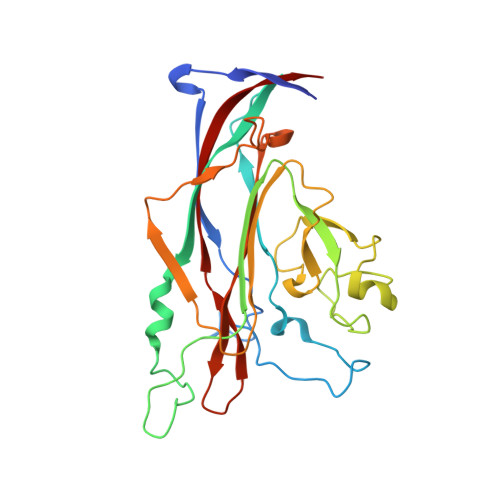
    3NXD, 3NXG

  • PubMed Abstract: 

    The human JC polyomavirus (JCV) causes a fatal demyelinating disease, progressive multifocal leukoencephalopathy (PML), in immunocompromised individuals. Current treatment options for PML are inadequate. Sialylated oligosaccharides and the serotonin receptor are known to be necessary for JCV entry, but the molecular interactions underlying JCV attachment remain unknown. Using glycan array screening and viral infectivity assays, we identify a linear sialylated pentasaccharide with the sequence NeuNAc-α2,6-Gal-β1,4-GlcNAc-β1,3-Gal-β1,4-Glc (LSTc) present on host glycoproteins and glycolipids as a specific JCV recognition motif. The crystal structure of the JCV capsid protein VP1 was solved alone and in complex with LSTc. It reveals extensive interactions with the terminal sialic acid of the LSTc motif and specific recognition of an extended conformation of LSTc. Mutations in the JCV oligosaccharide-binding sites abolish cell attachment, viral spread, and infectivity, further validating the importance of this interaction. Our findings provide a powerful platform for the development of antiviral compounds.


  • Organizational Affiliation
    • Interfaculty Institute for Biochemistry, University of Tübingen, D-72076 Tübingen, Germany.

Macromolecule Content 

  • Total Structure Weight: 151.15 kDa 
  • Atom Count: 11,187 
  • Modeled Residue Count: 1,291 
  • Deposited Residue Count: 1,360 
  • Unique protein chains: 1

Macromolecules

Find similar proteins by:|  3D Structure
Entity ID: 1
MoleculeChains  Sequence LengthOrganismDetailsImage
Major capsid protein VP1
A, B, C, D, E
272JC polyomavirusMutation(s): 0 
Gene Names: VP1
UniProt
Find proteins for P03089 (JC polyomavirus)
Explore P03089 
Go to UniProtKB:  P03089
Entity Groups
Sequence Clusters30% Identity50% Identity70% Identity90% Identity95% Identity100% Identity
UniProt GroupP03089
Sequence Annotations
Expand
Reference Sequence

Small Molecules

Ligands 2 Unique
IDChains Name / Formula / InChI Key2D Diagram3D Interactions
GOL

Query on GOL


Download:Ideal Coordinates CCD File
F [auth A],
I [auth B],
J [auth C],
L [auth D],
N [auth E]
GLYCEROL
C3 H8 O3
PEDCQBHIVMGVHV-UHFFFAOYSA-N
EDO

Query on EDO


Download:Ideal Coordinates CCD File
G [auth A],
H [auth A],
K [auth C],
M [auth D],
O [auth E]
1,2-ETHANEDIOL
C2 H6 O2
LYCAIKOWRPUZTN-UHFFFAOYSA-N

Experimental Data & Validation

Experimental Data

  • Method: X-RAY DIFFRACTION
  • Resolution: 1.95 Å
  • R-Value Free:  0.207 (Depositor), 0.208 (DCC) 
  • R-Value Work:  0.174 (Depositor), 0.175 (DCC) 
  • R-Value Observed: 0.175 (Depositor) 
Space Group: C 1 2 1
Unit Cell:
Length ( Å )Angle ( ˚ )
a = 149.57α = 90
b = 96.02β = 110.44
c = 128.55γ = 90
Software Package:
Software NamePurpose
RemDAqdata collection
PHASERphasing
REFMACrefinement
XDSdata reduction
XSCALEdata scaling

Structure Validation

View Full Validation Report


Entry History 

Deposition Data

Revision History  (Full details and data files)

  • Version 1.0: 2010-11-17
    Type: Initial release
  • Version 1.1: 2011-07-13
    Changes: Version format compliance
  • Version 1.2: 2024-02-21
    Changes: Data collection, Database references, Derived calculations, Refinement description