3NUG | pdb_00003nug

Crystal structure of wild type tetrameric pyridoxal 4-dehydrogenase from Mesorhizobium loti


Experimental Data Snapshot

  • Method: X-RAY DIFFRACTION
  • Resolution: 1.79 Å
  • R-Value Free: 
    0.202 (Depositor), 0.204 (DCC) 
  • R-Value Work: 
    0.152 (Depositor), 0.157 (DCC) 
  • R-Value Observed: 
    0.155 (Depositor) 

Starting Model: experimental
View more details

wwPDB Validation 3D Report Full Report

Validation slider image for 3NUG

Ligand Structure Quality Assessment 


This is version 1.3 of the entry. See complete history

Literature

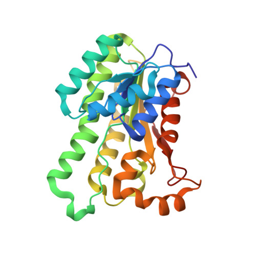
The crystal structure of SDR-type pyridoxal 4-dehydrogenase of Mesorhizobium loti

Chu, H.N.Kobayashi, J.Mikami, B.Yagi, T.

(2011) Biosci Biotechnol Biochem 75: 388-390

  • DOI: https://doi.org/10.1271/bbb.100748
  • Primary Citation Related Structures: 
    3NUG

  • PubMed Abstract: 

    Pyridoxal 4-dehydrogenase catalyzes the irreversible oxidation of pyridoxal to 4-pyridoxolactone and is involved in degradation pathway I of pyridoxine, a vitamin B(6) compound. Its crystal structure was elucidated for the first time. Molecular replacement with (S)-1-phenylthanol dehydrogenase (PDB code 2EW8) was adopted to determine the tertiary structure of the NAD(+)-bound enzyme.


  • Organizational Affiliation
    • Faculty of Agriculture and Agricultural Science Program, Graduate School of Integral Arts and Science, Kochi University, Nankoku, Kochi, Japan.

Macromolecule Content 

  • Total Structure Weight: 104.42 kDa 
  • Atom Count: 8,371 
  • Modeled Residue Count: 988 
  • Deposited Residue Count: 988 
  • Unique protein chains: 1

Macromolecules

Find similar proteins by:|  3D Structure
Entity ID: 1
MoleculeChains  Sequence LengthOrganismDetailsImage
3-oxoacyl-(Acyl-carrier protein) reductase
A, B, C, D
247Mesorhizobium lotiMutation(s): 0 
Gene Names: mlr6807pldh-t
EC: 1.1.1.107
UniProt
Find proteins for Q988B7 (Mesorhizobium japonicum (strain LMG 29417 / CECT 9101 / MAFF 303099))
Explore Q988B7 
Go to UniProtKB:  Q988B7
Entity Groups
Sequence Clusters30% Identity50% Identity70% Identity90% Identity95% Identity100% Identity
UniProt GroupQ988B7
Sequence Annotations
Expand
Reference Sequence

Experimental Data & Validation

Experimental Data

  • Method: X-RAY DIFFRACTION
  • Resolution: 1.79 Å
  • R-Value Free:  0.202 (Depositor), 0.204 (DCC) 
  • R-Value Work:  0.152 (Depositor), 0.157 (DCC) 
  • R-Value Observed: 0.155 (Depositor) 
Space Group: P 1 21 1
Unit Cell:
Length ( Å )Angle ( ˚ )
a = 85.638α = 90
b = 50.695β = 89.29
c = 94.551γ = 90
Software Package:
Software NamePurpose
HKL-2000data collection
PHASERphasing
PHENIXrefinement
HKL-2000data reduction
HKL-2000data scaling

Structure Validation

View Full Validation Report


Ligand Structure Quality Assessment 


Entry History 

Deposition Data

  • Released Date: 2011-04-20 
  • Deposition Author(s): Chu, H.N.

Revision History  (Full details and data files)

  • Version 1.0: 2011-04-20
    Type: Initial release
  • Version 1.1: 2011-07-13
    Changes: Version format compliance
  • Version 1.2: 2017-11-08
    Changes: Advisory, Refinement description
  • Version 1.3: 2023-11-01
    Changes: Advisory, Data collection, Database references, Derived calculations, Refinement description