3NSG | pdb_00003nsg

Crystal Structure of OmpF, an Outer Membrane Protein from Salmonella typhi


Experimental Data Snapshot

  • Method: X-RAY DIFFRACTION
  • Resolution: 2.79 Å
  • R-Value Free: 
    0.337 (Depositor), 0.350 (DCC) 
  • R-Value Work: 
    0.255 (Depositor), 0.281 (DCC) 
  • R-Value Observed: 
    0.259 (Depositor) 

wwPDB Validation 3D Report Full Report

Currently 3NSG does not have a validation slider image.


This is version 1.4 of the entry. See complete history

Literature

Asymmetric pore occupancy in crystal structure of OmpF porin from Salmonella typhi

Balasubramaniam, D.Arockiasamy, A.Kumar, P.D.Sharma, A.Krishnaswamy, S.

(2012) J Struct Biol 178: 233-244

  • DOI: https://doi.org/10.1016/j.jsb.2012.04.005
  • Primary Citation Related Structures: 
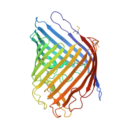
    3NSG

  • PubMed Abstract: 

    OmpF is a major general diffusion porin of Salmonella typhi, a Gram-negative bacterium, which is an obligatory human pathogen causing typhoid. The structure of S. typhi Ty21a OmpF (PDB Id: 3NSG) determined at 2.8 Å resolution by X-ray crystallography shows a 16-stranded β-barrel with three β-barrel monomers associated to form a trimer. The packing observed in S. typhi Ty21a rfOmpF crystals has not been observed earlier in other porin structures. The variations seen in the loop regions provide a starting point for using the S. typhi OmpF for structure-based multi-valent vaccine design. Along one side of the S. typhi Ty21a OmpF pore there exists a staircase arrangement of basic residues (20R, 60R, 62K, 65R, 77R, 130R and 16K), which also contribute, to the electrostatic potential in the pore. This structure suggests the presence of asymmetric electrostatics in the porin oligomer. Moreover, antibiotic translocation, permeability and reduced uptake in the case of mutants can be understood based on the structure paving the way for designing new antibiotics.


  • Organizational Affiliation
    • Department of Genetic Engineering, School of Biotechnology, Madurai Kamaraj University, Madurai 625 021, India.

Macromolecule Content 

  • Total Structure Weight: 131.77 kDa 
  • Atom Count: 9,432 
  • Modeled Residue Count: 1,023 
  • Deposited Residue Count: 1,023 
  • Unique protein chains: 1

Macromolecules

Find similar proteins by:|  3D Structure
Entity ID: 1
MoleculeChains  Sequence LengthOrganismDetailsImage
Outer membrane protein F
A, B, C
341Salmonella enterica subsp. enterica serovar Typhi str. Ty21aMutation(s): 0 
Gene Names: ompF
Membrane Entity: Yes 
UniProt
Find proteins for P37432 (Salmonella typhimurium (strain LT2 / SGSC1412 / ATCC 700720))
Explore P37432 
Go to UniProtKB:  P37432
Entity Groups
Sequence Clusters30% Identity50% Identity70% Identity90% Identity95% Identity100% Identity
UniProt GroupP37432
Sequence Annotations
Expand
Reference Sequence

Small Molecules

Ligands 6 Unique
IDChains Name / Formula / InChI Key2D Diagram3D Interactions
LDA

Query on LDA


Download:Ideal Coordinates CCD File
GE [auth C]
HE [auth C]
IE [auth C]
JC [auth B]
JE [auth C]
GE [auth C],
HE [auth C],
IE [auth C],
JC [auth B],
JE [auth C],
KC [auth B],
LC [auth B],
MC [auth B],
NC [auth B],
OC [auth B],
PC [auth B],
SA [auth A],
TA [auth A],
UA [auth A],
VA [auth A],
WA [auth A],
XA [auth A]
LAURYL DIMETHYLAMINE-N-OXIDE
C14 H31 N O
SYELZBGXAIXKHU-UHFFFAOYSA-N
FLC

Query on FLC


Download:Ideal Coordinates CCD File
AB [auth A],
ME [auth C],
NE [auth C],
QE [auth C]
CITRATE ANION
C6 H5 O7
KRKNYBCHXYNGOX-UHFFFAOYSA-K
TAM

Query on TAM


Download:Ideal Coordinates CCD File
KE [auth C]
LE [auth C]
QC [auth B]
SE [auth C]
YA [auth A]
KE [auth C],
LE [auth C],
QC [auth B],
SE [auth C],
YA [auth A],
ZA [auth A]
TRIS(HYDROXYETHYL)AMINOMETHANE
C7 H17 N O3
GKODZWOPPOTFGA-UHFFFAOYSA-N
TLA

Query on TLA


Download:Ideal Coordinates CCD File
BB [auth A],
RC [auth B]
L(+)-TARTARIC ACID
C4 H6 O6
FEWJPZIEWOKRBE-JCYAYHJZSA-N
SO4

Query on SO4


Download:Ideal Coordinates CCD File
AA [auth A]
AD [auth C]
BA [auth A]
BD [auth C]
CA [auth A]
AA [auth A],
AD [auth C],
BA [auth A],
BD [auth C],
CA [auth A],
CB [auth B],
CD [auth C],
D [auth A],
DA [auth A],
DB [auth B],
DD [auth C],
E [auth A],
EB [auth B],
ED [auth C],
F [auth A],
FB [auth B],
FD [auth C],
G [auth A],
GB [auth B],
GD [auth C],
H [auth A],
HB [auth B],
HD [auth C],
I [auth A],
IB [auth B],
ID [auth C],
J [auth A],
JB [auth B],
JD [auth C],
K [auth A],
KB [auth B],
KD [auth C],
L [auth A],
LB [auth B],
LD [auth C],
M [auth A],
MB [auth B],
MD [auth C],
N [auth A],
NB [auth B],
ND [auth C],
O [auth A],
OB [auth B],
OD [auth C],
P [auth A],
PB [auth B],
PD [auth C],
Q [auth A],
QB [auth B],
QD [auth C],
R [auth A],
RB [auth B],
RD [auth C],
S [auth A],
SB [auth B],
SC [auth C],
SD [auth C],
T [auth A],
TB [auth B],
TC [auth C],
TD [auth C],
U [auth A],
UB [auth B],
UC [auth C],
UD [auth C],
V [auth A],
VB [auth B],
VC [auth C],
VD [auth C],
W [auth A],
WB [auth B],
WC [auth C],
WD [auth C],
X [auth A],
XB [auth B],
XC [auth C],
Y [auth A],
YC [auth C],
Z [auth A],
ZC [auth C]
SULFATE ION
O4 S
QAOWNCQODCNURD-UHFFFAOYSA-L
GOL

Query on GOL


Download:Ideal Coordinates CCD File
AC [auth B]
AE [auth C]
BC [auth B]
BE [auth C]
CC [auth B]
AC [auth B],
AE [auth C],
BC [auth B],
BE [auth C],
CC [auth B],
CE [auth C],
DC [auth B],
DE [auth C],
EA [auth A],
EC [auth B],
EE [auth C],
FA [auth A],
FC [auth B],
FE [auth C],
GA [auth A],
GC [auth B],
HA [auth A],
HC [auth B],
IA [auth A],
IC [auth B],
JA [auth A],
KA [auth A],
LA [auth A],
MA [auth A],
NA [auth A],
OA [auth A],
OE [auth C],
PA [auth A],
PE [auth C],
QA [auth A],
RA [auth A],
RE [auth C],
TE [auth C],
XD [auth C],
YB [auth B],
YD [auth C],
ZB [auth B],
ZD [auth C]
GLYCEROL
C3 H8 O3
PEDCQBHIVMGVHV-UHFFFAOYSA-N
Modified Residues  1 Unique
IDChains TypeFormula2D DiagramParent
MSE
Query on MSE
A, B, C
L-PEPTIDE LINKINGC5 H11 N O2 SeMET

Experimental Data & Validation

Experimental Data

  • Method: X-RAY DIFFRACTION
  • Resolution: 2.79 Å
  • R-Value Free:  0.337 (Depositor), 0.350 (DCC) 
  • R-Value Work:  0.255 (Depositor), 0.281 (DCC) 
  • R-Value Observed: 0.259 (Depositor) 
Space Group: P 21 21 21
Unit Cell:
Length ( Å )Angle ( ˚ )
a = 85.185α = 90
b = 139.335β = 90
c = 150.772γ = 90
Software Package:
Software NamePurpose
DENZOdata reduction
SCALEPACKdata scaling
REFMACrefinement
PDB_EXTRACTdata extraction

Structure Validation

View Full Validation Report

Currently 3NSG does not have a validation slider image.


Entry History 

Deposition Data

Revision History  (Full details and data files)

  • Version 1.0: 2011-07-13
    Type: Initial release
  • Version 1.1: 2012-06-20
    Changes: Database references
  • Version 1.2: 2017-11-08
    Changes: Refinement description
  • Version 1.3: 2023-12-27
    Changes: Data collection, Database references, Derived calculations
  • Version 1.4: 2024-10-30
    Changes: Structure summary