Structural and biochemical studies of the open state of Lys48-linked diubiquitin.
Lai, M.Y., Zhang, D., Laronde-Leblanc, N., Fushman, D.(2012) Biochim Biophys Acta 1823: 2046-2056
- PubMed: 22542781 Search on PubMedSearch on PubMed Central
- DOI: https://doi.org/10.1016/j.bbamcr.2012.04.003
- Primary Citation Related Structures:
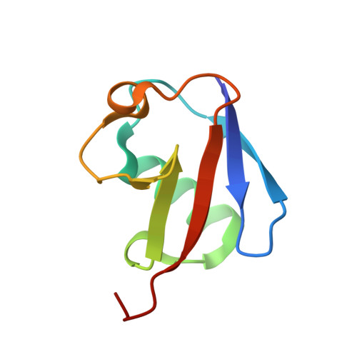
3NS8 - PubMed Abstract:
Ubiquitin (Ub) is a small protein highly conserved among eukaryotes and involved in practically all aspects of eukaryotic cell biology. Polymeric chains assembled from covalently-linked Ub monomers function as molecular signals in the regulation of a host of cellular processes. Our previous studies have shown that the predominant state of Lys48-linked di- and tetra-Ub chains at near-physiological conditions is a closed conformation, in which the Ub-Ub interface is formed by the hydrophobic surface residues of the adjacent Ub units. Because these very residues are involved in (poly)Ub interactions with the majority of Ub-binding proteins, their sequestration at the Ub-Ub interface renders the closed conformation of polyUb binding incompetent. Thus the existence of open conformation(s) and the interdomain motions opening and closing the Ub-Ub interface is critical for the recognition of Lys48-linked polyUb by its receptors. Knowledge of the conformational properties of a polyUb signal is essential for our understanding of its specific recognition by various Ub-receptors. Despite their functional importance, open states of Lys48-linked chains are poorly characterized. Here we report a crystal structure of the open state of Lys48-linked di-Ub. Moreover, using NMR, we examined interactions of the open state of this chain (at pH4.5) with a Lys48-linkage-selective receptor, the UBA2 domain of a shuttle protein hHR23a. Our results show that di-Ub binds UBA2 in the same mode and with comparable affinity as the closed state. Our data suggest a mechanism for polyUb signal recognition, whereby Ub-binding proteins select specific conformations out of the available ensemble of polyUb chain conformations. This article is part of a Special Issue entitled: Ubiquitin Drug Discovery and Diagnostics.
- Department of Chemistry and Biochemistry, University of Maryland, College Park, MD 20742, USA.
Organizational Affiliation: