3NMJ | pdb_00003nmj

Crystal structure of a nickel mediated dimer for the phenanthroline-modified cytochrome cb562 variant, MBP-Phen2


Experimental Data Snapshot

  • Method: X-RAY DIFFRACTION
  • Resolution: 3.10 Å
  • R-Value Free: 
    0.238 (Depositor), 0.234 (DCC) 
  • R-Value Work: 
    0.193 (Depositor), 0.190 (DCC) 
  • R-Value Observed: 
    0.196 (Depositor) 

wwPDB Validation 3D Report Full Report

Validation slider image for 3NMJ

Ligand Structure Quality Assessment 


This is version 1.2 of the entry. See complete history

Literature

Porous protein frameworks with unsaturated metal centers in sterically encumbered coordination sites.

Radford, R.J.Lawrenz, M.Nguyen, P.C.McCammon, J.A.Tezcan, F.A.

(2011) Chem Commun (Camb) 47: 313-315

  • DOI: https://doi.org/10.1039/c0cc02168g
  • Primary Citation Related Structures: 
    3NMI, 3NMJ, 3NMK

  • PubMed Abstract: 

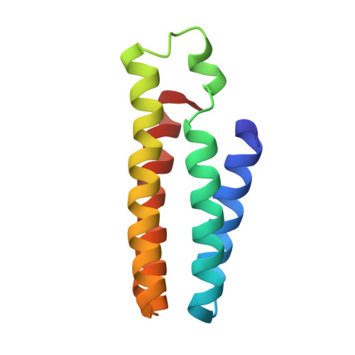
    Described is an engineered metal-binding protein, MBPPhen2, which forms porous crystalline frameworks that feature coordinatively unsaturated Zn- and Ni-centers.


  • Organizational Affiliation
    • Department of Chemistry and Biochemistry, University of California, San Diego 9500 Gilman Dr, La Jolla, CA 92037, USA.

Macromolecule Content 

  • Total Structure Weight: 49.38 kDa 
  • Atom Count: 3,437 
  • Modeled Residue Count: 423 
  • Deposited Residue Count: 424 
  • Unique protein chains: 1

Macromolecules

Find similar proteins by:|  3D Structure
Entity ID: 1
MoleculeChains  Sequence LengthOrganismDetailsImage
Soluble cytochrome b562
A, B, C, D
106Escherichia coliMutation(s): 6 
Gene Names: cybC
UniProt
Find proteins for P0ABE7 (Escherichia coli)
Explore P0ABE7 
Go to UniProtKB:  P0ABE7
Entity Groups
Sequence Clusters30% Identity50% Identity70% Identity90% Identity95% Identity100% Identity
UniProt GroupP0ABE7
Sequence Annotations
Expand
Reference Sequence

Small Molecules

Ligands 3 Unique
IDChains Name / Formula / InChI Key2D Diagram3D Interactions
HEM

Query on HEM


Download:Ideal Coordinates CCD File
E [auth A],
G [auth B],
K [auth C],
M [auth D]
PROTOPORPHYRIN IX CONTAINING FE
C34 H32 Fe N4 O4
KABFMIBPWCXCRK-RGGAHWMASA-L
PXX

Query on PXX


Download:Ideal Coordinates CCD File
F [auth A],
H [auth B],
L [auth C],
N [auth D]
N-1,10-phenanthrolin-5-ylacetamide
C14 H11 N3 O
AAJXINSCZMZERD-UHFFFAOYSA-N
NI

Query on NI


Download:Ideal Coordinates CCD File
I [auth B],
J [auth C]
NICKEL (II) ION
Ni
VEQPNABPJHWNSG-UHFFFAOYSA-N

Experimental Data & Validation

Experimental Data

  • Method: X-RAY DIFFRACTION
  • Resolution: 3.10 Å
  • R-Value Free:  0.238 (Depositor), 0.234 (DCC) 
  • R-Value Work:  0.193 (Depositor), 0.190 (DCC) 
  • R-Value Observed: 0.196 (Depositor) 
Space Group: P 65
Unit Cell:
Length ( Å )Angle ( ˚ )
a = 99.291α = 90
b = 99.291β = 90
c = 109.317γ = 120
Software Package:
Software NamePurpose
SCALAdata scaling
PHASERphasing
REFMACrefinement
PDB_EXTRACTdata extraction
Blu-Icedata collection
MOSFLMdata reduction

Structure Validation

View Full Validation Report


Ligand Structure Quality Assessment 


Entry History 

Deposition Data

Revision History  (Full details and data files)

  • Version 1.0: 2010-11-24
    Type: Initial release
  • Version 1.1: 2011-07-13
    Changes: Version format compliance
  • Version 1.2: 2023-12-27
    Changes: Data collection, Database references, Derived calculations