3NJW | pdb_00003njw

First High Resolution Crystal Structure of a Lasso Peptide


Experimental Data Snapshot

  • Method: X-RAY DIFFRACTION
  • Resolution: 0.86 Å
  • R-Value Observed: 
    0.094 (Depositor) 

wwPDB Validation 3D Report Full Report

Validation slider image for 3NJW

This is version 1.3 of the entry. See complete history

Literature

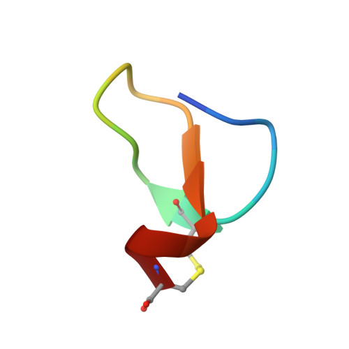
High-resolution crystal structure of a lasso Peptide.

Nar, H.Schmid, A.Puder, C.Potterat, O.

(2010) ChemMedChem 5: 1689-1692

Macromolecule Content 

  • Total Structure Weight: 2.06 kDa 
  • Atom Count: 169 
  • Modeled Residue Count: 19 
  • Deposited Residue Count: 19 
  • Unique protein chains: 1

Macromolecules

Find similar proteins by:  Sequence   |   3D Structure  
Entity ID: 1
MoleculeChains  Sequence LengthOrganismDetailsImage
Bicyclic peptide BI-3216919Streptomyces sp.Mutation(s): 0 

Experimental Data & Validation

Experimental Data

  • Method: X-RAY DIFFRACTION
  • Resolution: 0.86 Å
  • R-Value Observed: 0.094 (Depositor) 
Space Group: P 21 21 21
Unit Cell:
Length ( Å )Angle ( ˚ )
a = 19.465α = 90
b = 21.432β = 90
c = 29.523γ = 90
Software Package:
Software NamePurpose
StructureStudiodata collection
SHELXDphasing
SHELXL-97refinement
d*TREKdata reduction
d*TREKdata scaling

Structure Validation

View Full Validation Report


Entry History 

Deposition Data

Revision History  (Full details and data files)

  • Version 1.0: 2010-09-01
    Type: Initial release
  • Version 1.1: 2011-07-13
    Changes: Version format compliance
  • Version 1.2: 2011-08-10
    Changes: Atomic model, Database references
  • Version 1.3: 2024-11-20
    Changes: Data collection, Database references, Derived calculations, Structure summary