3NJG | pdb_00003njg

K98A mutant of SO1698 protein, an aspartic peptidase from Shewanella oneidensis.


Experimental Data Snapshot

  • Method: X-RAY DIFFRACTION
  • Resolution: 1.92 Å
  • R-Value Free: 
    0.196 (Depositor), 0.214 (DCC) 
  • R-Value Work: 
    0.168 (Depositor), 0.183 (DCC) 
  • R-Value Observed: 
    0.170 (Depositor) 

Starting Model: experimental
View more details

wwPDB Validation 3D Report Full Report

Validation slider image for 3NJG

This is version 1.4 of the entry. See complete history

Literature

Characterization of member of DUF1888 protein family, self-cleaving and self-assembling endopeptidase.

Osipiuk, J.Mulligan, R.Bargassa, M.Hamilton, J.E.Cunningham, M.A.Joachimiak, A.

(2012) J Biological Chem 287: 19452-19461

  • DOI: https://doi.org/10.1074/jbc.M112.358069
  • Primary Citation Related Structures: 
    3N55, 3NJF, 3NJG, 3NJH, 3NJI, 3NJJ, 3NJK, 3NJL, 3NJM, 3NJN

  • PubMed Abstract: 

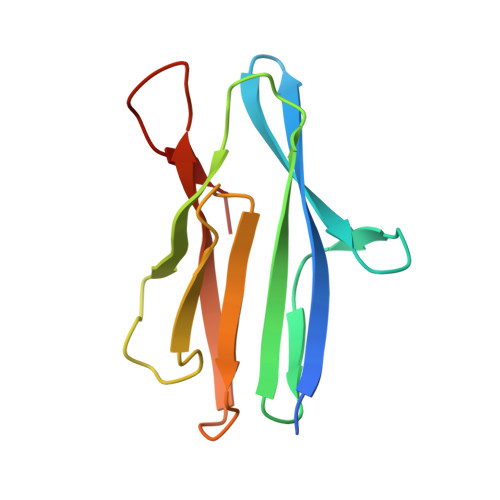
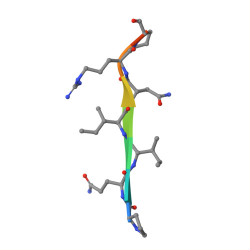
    The crystal structure of SO1698 protein from Shewanella oneidensis was determined by a SAD method and refined to 1.57 Å. The structure is a β sandwich that unexpectedly consists of two polypeptides; the N-terminal fragment includes residues 1-116, and the C-terminal one includes residues 117-125. Electron density also displayed the Lys-98 side chain covalently linked to Asp-116. The putative active site residues involved in self-cleavage were identified; point mutants were produced and characterized structurally and in a biochemical assay. Numerical simulations utilizing molecular dynamics and hybrid quantum/classical calculations suggest a mechanism involving activation of a water molecule coordinated by a catalytic aspartic acid.


  • Organizational Affiliation
    • Midwest Center for Structural Genomics and Structural Biology Center, Biosciences, Argonne National Laboratory, Argonne, Illinois 60439, USA.

Macromolecule Content 

  • Total Structure Weight: 13.69 kDa 
  • Atom Count: 1,045 
  • Modeled Residue Count: 119 
  • Deposited Residue Count: 128 
  • Unique protein chains: 2

Macromolecules

Find similar proteins by:|  3D Structure
Entity ID: 1
MoleculeChains  Sequence LengthOrganismDetailsImage
Peptidase119Shewanella oneidensisMutation(s): 1 
Gene Names: SO_1698
UniProt
Find proteins for Q8EGA7 (Shewanella oneidensis (strain ATCC 700550 / JCM 31522 / CIP 106686 / LMG 19005 / NCIMB 14063 / MR-1))
Explore Q8EGA7 
Go to UniProtKB:  Q8EGA7
Entity Groups
Sequence Clusters30% Identity50% Identity70% Identity90% Identity95% Identity100% Identity
UniProt GroupQ8EGA7
Sequence Annotations
Expand
Reference Sequence
Find similar proteins by:  Sequence   |   3D Structure  
Entity ID: 2
MoleculeChains  Sequence LengthOrganismDetailsImage
Peptidase9Shewanella oneidensisMutation(s): 0 
Gene Names: SO_1698
UniProt
Find proteins for Q8EGA7 (Shewanella oneidensis (strain ATCC 700550 / JCM 31522 / CIP 106686 / LMG 19005 / NCIMB 14063 / MR-1))
Explore Q8EGA7 
Go to UniProtKB:  Q8EGA7
Entity Groups
UniProt GroupQ8EGA7
Sequence Annotations
Expand
Reference Sequence

Experimental Data & Validation

Experimental Data

  • Method: X-RAY DIFFRACTION
  • Resolution: 1.92 Å
  • R-Value Free:  0.196 (Depositor), 0.214 (DCC) 
  • R-Value Work:  0.168 (Depositor), 0.183 (DCC) 
  • R-Value Observed: 0.170 (Depositor) 
Space Group: H 3 2
Unit Cell:
Length ( Å )Angle ( ˚ )
a = 99.901α = 90
b = 99.901β = 90
c = 100.589γ = 120
Software Package:
Software NamePurpose
DENZOdata reduction
SCALEPACKdata scaling
MOLREPphasing
REFMACrefinement
PDB_EXTRACTdata extraction
SBC-Collectdata collection
HKL-2000data reduction
HKL-2000data scaling

Structure Validation

View Full Validation Report


Entry History 

Deposition Data

Revision History  (Full details and data files)

  • Version 1.0: 2010-07-14
    Type: Initial release
  • Version 1.1: 2011-07-13
    Changes: Version format compliance
  • Version 1.2: 2012-10-10
    Changes: Database references
  • Version 1.3: 2017-11-08
    Changes: Refinement description
  • Version 1.4: 2023-09-06
    Changes: Data collection, Database references, Refinement description