3NIO | pdb_00003nio

Crystal structure of Pseudomonas aeruginosa guanidinobutyrase


Experimental Data Snapshot

  • Method: X-RAY DIFFRACTION
  • Resolution: 2.00 Å
  • R-Value Free: 
    0.251 (Depositor), 0.267 (DCC) 
  • R-Value Work: 
    0.233 (Depositor), 0.251 (DCC) 
  • R-Value Observed: 
    0.234 (Depositor) 

Starting Model: experimental
View more details

wwPDB Validation 3D Report Full Report

Validation slider image for 3NIO

This is version 1.3 of the entry. See complete history

Literature

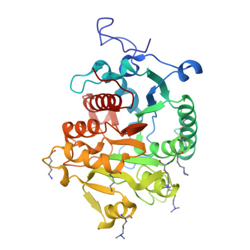
Crystal structures of Pseudomonas aeruginosa guanidinobutyrase and guanidinopropionase, members of the ureohydrolase superfamily

Lee, S.J.Kim, D.J.Kim, H.S.Lee, B.I.Yoon, H.J.Yoon, J.Y.Kim, K.H.Jang, J.Y.Im, H.N.An, D.R.Song, J.S.Kim, H.J.Suh, S.W.

(2011) J Struct Biol 175: 329-338

  • DOI: https://doi.org/10.1016/j.jsb.2011.05.002
  • Primary Citation Related Structures: 
    3NIO, 3NIP, 3NIQ

  • PubMed Abstract: 

    Pseudomonas aeruginosa guanidinobutyrase (GbuA) and guanidinopropionase (GpuA) catalyze the hydrolysis of 4-guanidinobutyrate and 3-guanidinopropionate, respectively. They belong to the ureohydrolase superfamily, which includes arginase, agmatinase, proclavaminate amidinohydrolase, and formiminoglutamase. In this study, we have determined the crystal structures of GbuA and GpuA from P. aeruginosa to provide a structural insight into their substrate specificity. Although GbuA and GpuA share a common structural fold of the typical ureohydrolase superfamily, they exhibit significant variations in two active site loops. Mutagenesis of Met161 of GbuA and Tyr157 of GpuA, both of which are located in the active site loop 1 and predicted to be involved in substrate recognition, significantly affected their enzymatic properties, implying their important roles in catalysis.


  • Organizational Affiliation
    • Department of Chemistry, College of Natural Sciences, Seoul National University, Seoul 151-742, Republic of Korea.

Macromolecule Content 

  • Total Structure Weight: 210.4 kDa 
  • Atom Count: 15,530 
  • Modeled Residue Count: 1,896 
  • Deposited Residue Count: 1,914 
  • Unique protein chains: 1

Macromolecules

Find similar proteins by:|  3D Structure
Entity ID: 1
MoleculeChains  Sequence LengthOrganismDetailsImage
Guanidinobutyrase
A, B, C, D, E
A, B, C, D, E, F
319Pseudomonas aeruginosa PAO1Mutation(s): 0 
Gene Names: gbuAPA1421
EC: 3.5.3.7
UniProt
Find proteins for Q9I3S3 (Pseudomonas aeruginosa (strain ATCC 15692 / DSM 22644 / CIP 104116 / JCM 14847 / LMG 12228 / 1C / PRS 101 / PAO1))
Explore Q9I3S3 
Go to UniProtKB:  Q9I3S3
Entity Groups
Sequence Clusters30% Identity50% Identity70% Identity90% Identity95% Identity100% Identity
UniProt GroupQ9I3S3
Sequence Annotations
Expand
Reference Sequence

Small Molecules

Ligands 1 Unique
IDChains Name / Formula / InChI Key2D Diagram3D Interactions
MN

Query on MN


Download:Ideal Coordinates CCD File
G [auth A]
H [auth A]
I [auth B]
J [auth B]
K [auth C]
G [auth A],
H [auth A],
I [auth B],
J [auth B],
K [auth C],
L [auth C],
M [auth D],
N [auth D],
O [auth E],
P [auth E],
Q [auth F],
R [auth F]
MANGANESE (II) ION
Mn
WAEMQWOKJMHJLA-UHFFFAOYSA-N
Modified Residues  1 Unique
IDChains TypeFormula2D DiagramParent
MLY
Query on MLY
A, B, C, D, E
A, B, C, D, E, F
L-PEPTIDE LINKINGC8 H18 N2 O2LYS

Experimental Data & Validation

Experimental Data

  • Method: X-RAY DIFFRACTION
  • Resolution: 2.00 Å
  • R-Value Free:  0.251 (Depositor), 0.267 (DCC) 
  • R-Value Work:  0.233 (Depositor), 0.251 (DCC) 
  • R-Value Observed: 0.234 (Depositor) 
Space Group: P 1 21 1
Unit Cell:
Length ( Å )Angle ( ˚ )
a = 82.047α = 90
b = 158.954β = 114.84
c = 84.547γ = 90
Software Package:
Software NamePurpose
HKL-2000data collection
CNSrefinement
REFMACrefinement
HKL-2000data reduction
HKL-2000data scaling
CNSphasing

Structure Validation

View Full Validation Report


Entry History 

Deposition Data

Revision History  (Full details and data files)

  • Version 1.0: 2011-06-01
    Type: Initial release
  • Version 1.1: 2011-07-13
    Changes: Version format compliance
  • Version 1.2: 2014-02-26
    Changes: Database references
  • Version 1.3: 2023-11-01
    Changes: Data collection, Database references, Derived calculations, Refinement description