3NH0 | pdb_00003nh0

Crystal structure of RNase T in complex with a non-preferred ssDNA (AAC)


Experimental Data Snapshot

  • Method: X-RAY DIFFRACTION
  • Resolution: 2.30 Å
  • R-Value Free: 
    0.249 (Depositor), 0.247 (DCC) 
  • R-Value Work: 
    0.196 (Depositor), 0.193 (DCC) 
  • R-Value Observed: 
    0.200 (Depositor) 

Starting Model: experimental
View more details

wwPDB Validation 3D Report Full Report

Validation slider image for 3NH0

This is version 1.3 of the entry. See complete history

Literature

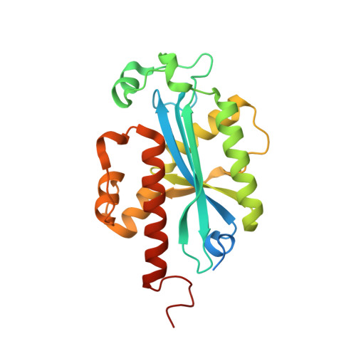
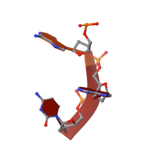
Structural basis for RNA trimming by RNase T in stable RNA 3'-end maturation

Hsiao, Y.-Y.Yang, C.-C.Lin, C.L.Lin, J.L.J.Duh, Y.Yuan, H.S.

(2011) Nat Chem Biol 7: 236-243

  • DOI: https://doi.org/10.1038/nchembio.524
  • Primary Citation Related Structures: 
    3NGY, 3NGZ, 3NH0, 3NH1, 3NH2

  • PubMed Abstract: 

    RNA maturation relies on various exonucleases to remove nucleotides successively from the 5' or 3' end of nucleic acids. However, little is known regarding the molecular basis for substrate and cleavage preference of exonucleases. Our biochemical and structural analyses on RNase T-DNA complexes show that the RNase T dimer has an ideal architecture for binding a duplex with a short 3' overhang to produce a digestion product of a duplex with a 2-nucleotide (nt) or 1-nt 3' overhang, depending on the composition of the last base pair in the duplex. A 'C-filter' in RNase T screens out the nucleic acids with 3'-terminal cytosines for hydrolysis by inducing a disruptive conformational change at the active site. Our results reveal the general principles and the working mechanism for the final trimming step made by RNase T in the maturation of stable RNA and pave the way for the understanding of other DEDD family exonucleases.


  • Organizational Affiliation
    • Institute of Bioinformatics and Structural Biology, National Tsing Hua University, Hsinchu, Taiwan, ROC.

Macromolecule Content 

  • Total Structure Weight: 55.6 kDa 
  • Atom Count: 3,576 
  • Modeled Residue Count: 420 
  • Deposited Residue Count: 484 
  • Unique protein chains: 1
  • Unique nucleic acid chains: 1

Macromolecules


Find similar proteins by:|  3D Structure
Entity ID: 1
MoleculeChains  Sequence LengthOrganismDetailsImage
Ribonuclease T
A, B
235Escherichia coliMutation(s): 0 
Gene Names: rnt
EC: 3.1.13
UniProt
Find proteins for P30014 (Escherichia coli (strain K12))
Explore P30014 
Go to UniProtKB:  P30014
Entity Groups
Sequence Clusters30% Identity50% Identity70% Identity90% Identity95% Identity100% Identity
UniProt GroupP30014
Sequence Annotations
Expand
Reference Sequence
Find similar nucleic acids by:  Sequence
Entity ID: 2
MoleculeChains LengthOrganismImage
5'-D(*TP*TP*AP*CP*AP*AP*C)-3'
C, D
7N/A
Sequence Annotations
Expand
Reference Sequence

Experimental Data & Validation

Experimental Data

  • Method: X-RAY DIFFRACTION
  • Resolution: 2.30 Å
  • R-Value Free:  0.249 (Depositor), 0.247 (DCC) 
  • R-Value Work:  0.196 (Depositor), 0.193 (DCC) 
  • R-Value Observed: 0.200 (Depositor) 
Space Group: P 21 21 2
Unit Cell:
Length ( Å )Angle ( ˚ )
a = 96.075α = 90
b = 106.36β = 90
c = 46.991γ = 90
Software Package:
Software NamePurpose
HKL-2000data collection
AMoREphasing
PHENIXrefinement
HKL-2000data reduction
HKL-2000data scaling

Structure Validation

View Full Validation Report


Entry History 

Deposition Data

Revision History  (Full details and data files)

  • Version 1.0: 2011-02-16
    Type: Initial release
  • Version 1.1: 2011-07-13
    Changes: Version format compliance
  • Version 1.2: 2011-12-21
    Changes: Database references
  • Version 1.3: 2023-11-01
    Changes: Data collection, Database references, Refinement description