The function and three-dimensional structure of a thromboxane a(2)/cysteinyl leukotriene-binding protein from the saliva of a mosquito vector of the malaria parasite.
Alvarenga, P.H., Francischetti, I.M., Calvo, E., Sa-Nunes, A., Ribeiro, J.M., Andersen, J.F.(2010) PLoS Biol 8: e1000547-e1000547
- PubMed: 21152418 Search on PubMedSearch on PubMed Central
- DOI: https://doi.org/10.1371/journal.pbio.1000547
- Primary Citation Related Structures:
3NGV, 3NHI, 3NHT - PubMed Abstract:
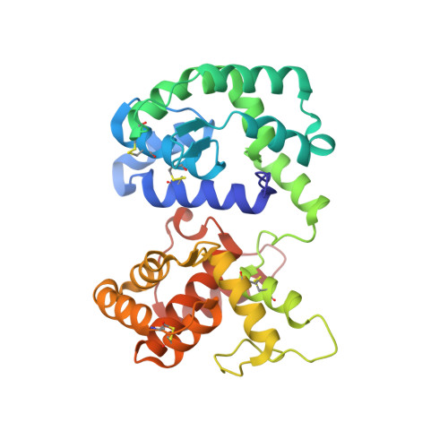
The highly expressed D7 protein family of mosquito saliva has previously been shown to act as an anti-inflammatory mediator by binding host biogenic amines and cysteinyl leukotrienes (CysLTs). In this study we demonstrate that AnSt-D7L1, a two-domain member of this group from Anopheles stephensi, retains the CysLT binding function seen in the homolog AeD7 from Aedes aegypti but has lost the ability to bind biogenic amines. Unlike any previously characterized members of the D7 family, AnSt-D7L1 has acquired the important function of binding thromboxane A(2) (TXA(2)) and its analogs with high affinity. When administered to tissue preparations, AnSt-D7L1 abrogated Leukotriene C(4) (LTC(4))-induced contraction of guinea pig ileum and contraction of rat aorta by the TXA(2) analog U46619. The protein also inhibited platelet aggregation induced by both collagen and U46619 when administered to stirred platelets. The crystal structure of AnSt-D7L1 contains two OBP-like domains and has a structure similar to AeD7. In AnSt-D7L1, the binding pocket of the C-terminal domain has been rearranged relative to AeD7, making the protein unable to bind biogenic amines. Structures of the ligand complexes show that CysLTs and TXA(2) analogs both bind in the same hydrophobic pocket of the N-terminal domain. The TXA(2) analog U46619 is stabilized by hydrogen bonding interactions of the ω-5 hydroxyl group with the phenolic hydroxyl group of Tyr 52. LTC(4) and occupies a very similar position to LTE(4) in the previously determined structure of its complex with AeD7. As yet, it is not known what, if any, new function has been acquired by the rearranged C-terminal domain. This article presents, to our knowledge, the first structural characterization of a protein from mosquito saliva that inhibits collagen mediated platelet activation.
- Laboratory of Malaria and Vector Research, National Institutes of Health, National Institute of Allergy and Infectious Diseases, Rockville, Maryland, United States of America.
Organizational Affiliation: