3NGB | pdb_00003ngb

Crystal structure of broadly and potently neutralizing antibody VRC01 in complex with HIV-1 gp120


Experimental Data Snapshot

  • Method: X-RAY DIFFRACTION
  • Resolution: 2.68 Å
  • R-Value Free: 
    0.256 (Depositor), 0.248 (DCC) 
  • R-Value Work: 
    0.197 (Depositor), 0.189 (DCC) 
  • R-Value Observed: 
    0.200 (Depositor) 

Starting Model: experimental
View more details

wwPDB Validation 3D Report Full Report

Validation slider image for 3NGB

Ligand Structure Quality Assessment 


This is version 3.3 of the entry. See complete history

Literature

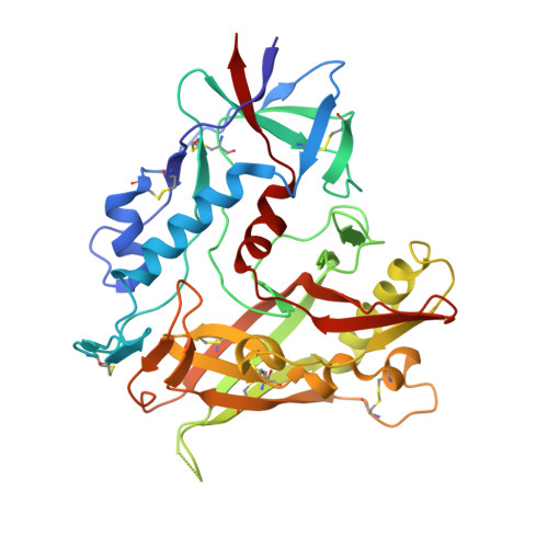
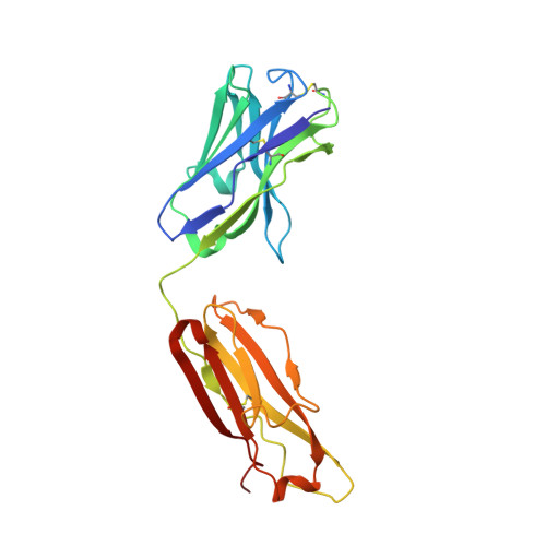
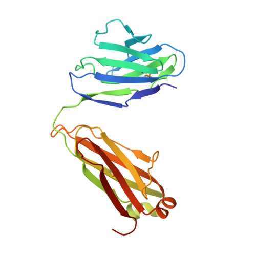
Structural basis for broad and potent neutralization of HIV-1 by antibody VRC01.

Zhou, T.Georgiev, I.Wu, X.Yang, Z.Y.Dai, K.Finzi, A.Do Kwon, Y.Scheid, J.F.Shi, W.Xu, L.Yang, Y.Zhu, J.Nussenzweig, M.C.Sodroski, J.Shapiro, L.Nabel, G.J.Mascola, J.R.Kwong, P.D.

(2010) Science 329: 811-817

  • DOI: https://doi.org/10.1126/science.1192819
  • Primary Citation Related Structures: 
    3NGB

  • PubMed Abstract: 

    During HIV-1 infection, antibodies are generated against the region of the viral gp120 envelope glycoprotein that binds CD4, the primary receptor for HIV-1. Among these antibodies, VRC01 achieves broad neutralization of diverse viral strains. We determined the crystal structure of VRC01 in complex with a human immunodeficiency virus HIV-1 gp120 core. VRC01 partially mimics CD4 interaction with gp120. A shift from the CD4-defined orientation, however, focuses VRC01 onto the vulnerable site of initial CD4 attachment, allowing it to overcome the glycan and conformational masking that diminishes the neutralization potency of most CD4-binding-site antibodies. To achieve this recognition, VRC01 contacts gp120 mainly through immunoglobulin V-gene regions substantially altered from their genomic precursors. Partial receptor mimicry and extensive affinity maturation thus facilitate neutralization of HIV-1 by natural human antibodies.


  • Organizational Affiliation
    • Vaccine Research Center, National Institute of Allergy and Infectious Diseases, National Institutes of Health (NIH), Bethesda, MD 20892, USA.

Macromolecule Content 

  • Total Structure Weight: 363.54 kDa 
  • Atom Count: 25,808 
  • Modeled Residue Count: 3,127 
  • Deposited Residue Count: 3,148 
  • Unique protein chains: 3

Macromolecules

Find similar proteins by:|  3D Structure
Entity ID: 1
MoleculeChains  Sequence LengthOrganismDetailsImage
Envelope glycoprotein gp160A [auth G],
D [auth A],
G [auth D],
J [auth I]
353Human immunodeficiency virus 1Mutation(s): 0 
Gene Names: Envpol
UniProt
Find proteins for Q0ED31 (Human immunodeficiency virus type 1)
Explore Q0ED31 
Go to UniProtKB:  Q0ED31
Entity Groups
Sequence Clusters30% Identity50% Identity70% Identity90% Identity95% Identity100% Identity
UniProt GroupQ0ED31
Glycosylation
Glycosylation Sites: 12
Sequence Annotations
Expand
Reference Sequence
Find similar proteins by:|  3D Structure
Entity ID: 2
MoleculeChains  Sequence LengthOrganismDetailsImage
Antigen binding fragment of heavy chain: Antibody VRC01B [auth H],
E [auth B],
H [auth E],
K [auth J]
224Homo sapiensMutation(s): 0 
Gene Names: Heavy chain
Find similar proteins by:|  3D Structure
Entity ID: 3
MoleculeChains  Sequence LengthOrganismDetailsImage
Antigen binding fragment of light chain: Antibody VRC01C [auth L],
F [auth C],
I [auth F],
L [auth K]
210Homo sapiensMutation(s): 0 
Gene Names: Light chain

Oligosaccharides

Help  
Entity ID: 4
MoleculeChains Length2D Diagram GlycosylationD Interactions
alpha-D-mannopyranose-(1-3)-[alpha-D-mannopyranose-(1-6)]beta-D-mannopyranose-(1-4)-2-acetamido-2-deoxy-beta-D-glucopyranose-(1-4)-2-acetamido-2-deoxy-beta-D-glucopyranose
M, N, P
5N-Glycosylation
Glycosylation Resources
GlyTouCan: G22768VO
GlyCosmos: G22768VO
GlyGen: G22768VO
Entity ID: 5
MoleculeChains Length2D Diagram GlycosylationD Interactions
2-acetamido-2-deoxy-beta-D-glucopyranose-(1-4)-2-acetamido-2-deoxy-beta-D-glucopyranose
O
2N-Glycosylation
Glycosylation Resources
GlyTouCan: G42666HT
GlyCosmos: G42666HT
GlyGen: G42666HT

Small Molecules

Ligands 3 Unique
IDChains Name / Formula / InChI Key2D Diagram3D Interactions
NAG

Query on NAG


Download:Ideal Coordinates CCD File
AA [auth G]
AB [auth D]
BB [auth D]
CA [auth A]
CB [auth D]
AA [auth G],
AB [auth D],
BB [auth D],
CA [auth A],
CB [auth D],
DA [auth A],
DB [auth D],
EA [auth A],
EB [auth D],
FA [auth A],
FB [auth D],
GA [auth A],
GB [auth D],
HA [auth A],
HB [auth D],
IA [auth A],
IB [auth D],
JA [auth A],
KA [auth A],
LA [auth A],
MA [auth A],
NA [auth A],
NB [auth I],
OB [auth I],
PB [auth I],
Q [auth G],
QB [auth I],
R [auth G],
RB [auth I],
S [auth G],
SB [auth I],
T [auth G],
TB [auth I],
U [auth G],
UB [auth I],
V [auth G],
VB [auth I],
W [auth G],
WB [auth I],
X [auth G],
XA [auth D],
XB [auth I],
Y [auth G],
YA [auth D],
Z [auth G],
ZA [auth D]
2-acetamido-2-deoxy-beta-D-glucopyranose
C8 H15 N O6
OVRNDRQMDRJTHS-FMDGEEDCSA-N
BGC

Query on BGC


Download:Ideal Coordinates CCD File
BA [auth G]
CC [auth J]
LB [auth D]
MB [auth E]
QA [auth A]
BA [auth G],
CC [auth J],
LB [auth D],
MB [auth E],
QA [auth A],
RA [auth A],
SA [auth A],
TA [auth A],
UA [auth B],
VA [auth B],
WA [auth C],
YB [auth I],
ZB [auth I]
beta-D-glucopyranose
C6 H12 O6
WQZGKKKJIJFFOK-VFUOTHLCSA-N
TRS

Query on TRS


Download:Ideal Coordinates CCD File
AC [auth I]
BC [auth J]
DC [auth K]
JB [auth D]
KB [auth D]
AC [auth I],
BC [auth J],
DC [auth K],
JB [auth D],
KB [auth D],
OA [auth A],
PA [auth A]
2-AMINO-2-HYDROXYMETHYL-PROPANE-1,3-DIOL
C4 H12 N O3
LENZDBCJOHFCAS-UHFFFAOYSA-O

Experimental Data & Validation

Experimental Data

  • Method: X-RAY DIFFRACTION
  • Resolution: 2.68 Å
  • R-Value Free:  0.256 (Depositor), 0.248 (DCC) 
  • R-Value Work:  0.197 (Depositor), 0.189 (DCC) 
  • R-Value Observed: 0.200 (Depositor) 
Space Group: P 1 21 1
Unit Cell:
Length ( Å )Angle ( ˚ )
a = 108.63α = 90
b = 98.275β = 99.68
c = 205.256γ = 90
Software Package:
Software NamePurpose
APSdata collection
PHASERphasing
PHENIXrefinement
HKL-2000data reduction
HKL-2000data scaling

Structure Validation

View Full Validation Report


Ligand Structure Quality Assessment 


Entry History 

Deposition Data

Revision History  (Full details and data files)

  • Version 1.0: 2010-07-07
    Type: Initial release
  • Version 1.1: 2011-07-13
    Changes: Version format compliance
  • Version 1.2: 2017-08-23
    Changes: Refinement description, Source and taxonomy
  • Version 2.0: 2020-07-29
    Type: Remediation
    Reason: Carbohydrate remediation
    Changes: Advisory, Atomic model, Data collection, Database references, Derived calculations, Structure summary
  • Version 3.0: 2020-10-14
    Changes: Advisory, Atomic model, Data collection, Derived calculations, Refinement description, Structure summary
  • Version 3.1: 2021-03-31
    Changes: Source and taxonomy
  • Version 3.2: 2023-09-06
    Changes: Data collection, Database references, Refinement description
  • Version 3.3: 2024-11-27
    Changes: Structure summary