


Crystal structure and inhibition of the beta-aminopeptidase BapA, a new ampicillin-recognizing member of the N-terminal nucleophile hydrolase family
Heck, T., Merz, T., Geueke, B., Gruetter, M.G., Kohler, H.-P.E.To be published.
Experimental Data Snapshot
Starting Model: experimental
View more details
Entity ID: 1 | |||||
|---|---|---|---|---|---|
| Molecule | Chains | Sequence Length | Organism | Details | Image |
| Beta-peptidyl aminopeptidase | 373 | Sphingosinicella xenopeptidilytica | Mutation(s): 0 Gene Names: bapA EC: 3.4.11.25 | 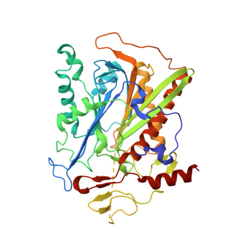 | |
UniProt | |||||
Entity Groups | |||||
| Sequence Clusters | 30% Identity50% Identity70% Identity90% Identity95% Identity100% Identity | ||||
| UniProt Group | Q52VH2 | ||||
Sequence AnnotationsExpand | |||||
Reference Sequence | |||||
| Ligands 3 Unique | |||||
|---|---|---|---|---|---|
| ID | Chains | Name / Formula / InChI Key | 2D Diagram | 3D Interactions | |
| OAE Download:Ideal Coordinates CCD File | G [auth A] H [auth A] J [auth B] N [auth C] P [auth D] | (2S,4S)-2-[(R)-{[(2R)-2-amino-2-phenylacetyl]amino}(carboxy)methyl]-5,5-dimethyl-1,3-thiazolidine-4-carboxylic acid C16 H21 N3 O5 S KDAWOPKDXRJNHV-BLFANLJRSA-N |  | ||
| SO4 Download:Ideal Coordinates CCD File | I [auth A], K [auth B], O [auth C], R [auth D] | SULFATE ION O4 S QAOWNCQODCNURD-UHFFFAOYSA-L |  | ||
| GOL Download:Ideal Coordinates CCD File | E [auth A], F [auth A], L [auth C], M [auth C] | GLYCEROL C3 H8 O3 PEDCQBHIVMGVHV-UHFFFAOYSA-N |  | ||
| Length ( Å ) | Angle ( ˚ ) |
|---|---|
| a = 88.3 | α = 90 |
| b = 97.1 | β = 108.7 |
| c = 102.2 | γ = 90 |
| Software Name | Purpose |
|---|---|
| XDS | data scaling |
| PHASER | phasing |
| PHENIX | refinement |
| XDS | data reduction |
| XSCALE | data scaling |


