


Investigation of 2-Fold Disorder of Inhibitors and Relative Potency by Crystallizations of HIV-1 Protease in Ritonavir and Saquinavir Mixtures
Olajuyigbe, F.M., Demitri, N., Geremia, S.(2011) Cryst Growth Des 11: 4378-4385
Experimental Data Snapshot
(2011) Cryst Growth Des 11: 4378-4385
Entity ID: 1 | |||||
|---|---|---|---|---|---|
| Molecule | Chains | Sequence Length | Organism | Details | Image |
| Protease | 99 | Human immunodeficiency virus 1 | Mutation(s): 5 Gene Names: pol EC: 3.4.23.16 | 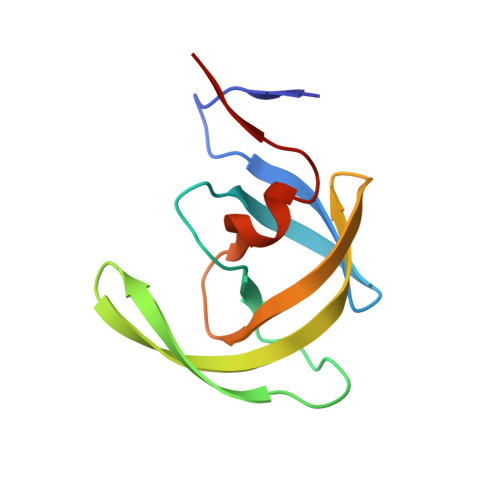 | |
UniProt | |||||
Entity Groups | |||||
| Sequence Clusters | 30% Identity50% Identity70% Identity90% Identity95% Identity100% Identity | ||||
| UniProt Group | P03367 | ||||
Sequence AnnotationsExpand | |||||
Reference Sequence | |||||
| Ligands 5 Unique | |||||
|---|---|---|---|---|---|
| ID | Chains | Name / Formula / InChI Key | 2D Diagram | 3D Interactions | |
| RIT Download:Ideal Coordinates CCD File | C [auth A] | RITONAVIR C37 H48 N6 O5 S2 NCDNCNXCDXHOMX-XGKFQTDJSA-N |  | ||
| SO4 Download:Ideal Coordinates CCD File | G [auth B], L [auth B] | SULFATE ION O4 S QAOWNCQODCNURD-UHFFFAOYSA-L |  | ||
| GOL Download:Ideal Coordinates CCD File | E [auth A], H [auth B] | GLYCEROL C3 H8 O3 PEDCQBHIVMGVHV-UHFFFAOYSA-N |  | ||
| DMS Download:Ideal Coordinates CCD File | D [auth A], I [auth B], J [auth B], K [auth B] | DIMETHYL SULFOXIDE C2 H6 O S IAZDPXIOMUYVGZ-UHFFFAOYSA-N |  | ||
| CL Download:Ideal Coordinates CCD File | F [auth A] | CHLORIDE ION Cl VEXZGXHMUGYJMC-UHFFFAOYSA-M |  | ||
| Entity ID: 2 | |||||
|---|---|---|---|---|---|
| ID | Chains | Name | Type/Class | 2D Diagram | 3D Interactions |
| PRD_001001 (RIT) Query on PRD_001001 | C [auth A] | RITONAVIR | Peptide-like / Inhibitor | | |
| Length ( Å ) | Angle ( ˚ ) |
|---|---|
| a = 51.16 | α = 90 |
| b = 58.567 | β = 90 |
| c = 61.143 | γ = 90 |
| Software Name | Purpose |
|---|---|
| HKL-2000 | data collection |
| SHELX | model building |
| SHELXL-97 | refinement |
| HKL-2000 | data reduction |
| HKL-2000 | data scaling |
| SHELX | phasing |


