3N8S | pdb_00003n8s

Crystal Structure of BlaC-E166A covalently bound with Cefamandole


Experimental Data Snapshot

  • Method: X-RAY DIFFRACTION
  • Resolution: 2.00 Å
  • R-Value Free: 
    0.204 (Depositor), 0.202 (DCC) 
  • R-Value Work: 
    0.161 (Depositor), 0.164 (DCC) 
  • R-Value Observed: 
    0.163 (Depositor) 

wwPDB Validation 3D Report Full Report

Validation slider image for 3N8S

Ligand Structure Quality Assessment 


This is version 1.4 of the entry. See complete history

Literature

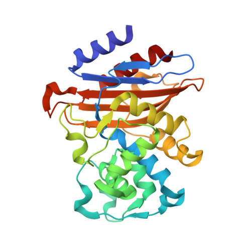
Structures of the Michaelis Complex (1.2 A) and the Covalent Acyl Intermediate (2.0 A) of Cefamandole Bound in the Active Sites of the Mycobacterium tuberculosis beta-Lactamase K73A and E166A Mutants.

Tremblay, L.W.Xu, H.Blanchard, J.S.

(2010) Biochemistry 49: 9685-9687

  • DOI: https://doi.org/10.1021/bi1015088
  • Primary Citation Related Structures: 
    3N8S, 3NY4

  • PubMed Abstract: 

    The genome of Mycobacterium tuberculosis (TB) contains a gene that encodes a highly active β-lactamase, BlaC, that imparts TB with resistance to β-lactam chemotherapy. The structure of covalent BlaC-β-lactam complexes suggests that active site residues K73 and E166 are essential for acylation and deacylation, respectively. We have prepared the K73A and E166A mutant forms of BlaC and have determined the structures of the Michaelis complex of cefamandole and the covalently bound acyl intermediate of cefamandole at resolutions of 1.2 and 2.0 Å, respectively. These structures provide insight into the details of the catalytic mechanism.


  • Organizational Affiliation
    • Department of Biochemistry, Albert Einstein College of Medicine, 1300 Morris Park Avenue, Bronx, New York 10461, United States.

Macromolecule Content 

  • Total Structure Weight: 28.87 kDa 
  • Atom Count: 2,311 
  • Modeled Residue Count: 265 
  • Deposited Residue Count: 265 
  • Unique protein chains: 1

Macromolecules

Find similar proteins by:|  3D Structure
Entity ID: 1
MoleculeChains  Sequence LengthOrganismDetailsImage
Beta-lactamase265Mycobacterium tuberculosisMutation(s): 1 
Gene Names: blaAblaCMT2128MTCY49.07cRv2068c
EC: 3.5.2.6
UniProt
Find proteins for P9WKD3 (Mycobacterium tuberculosis (strain ATCC 25618 / H37Rv))
Explore P9WKD3 
Go to UniProtKB:  P9WKD3
Entity Groups
Sequence Clusters30% Identity50% Identity70% Identity90% Identity95% Identity100% Identity
UniProt GroupP9WKD3
Sequence Annotations
Expand
Reference Sequence

Small Molecules

Ligands 2 Unique
IDChains Name / Formula / InChI Key2D Diagram3D Interactions
XD2

Query on XD2


Download:Ideal Coordinates CCD File
D [auth A](2R)-2-[(1R)-1-{[(2R)-2-hydroxy-2-phenylacetyl]amino}-2-oxoethyl]-5-{[(1-methyl-1H-tetrazol-5-yl)sulfanyl]methyl}-3,6-dihydro-2H-1,3-thiazine-4-carboxylic acid
C18 H20 N6 O5 S2
CTWPBVTUYYLUGC-XNRPHZJLSA-N
PO4

Query on PO4


Download:Ideal Coordinates CCD File
B [auth A],
C [auth A]
PHOSPHATE ION
O4 P
NBIIXXVUZAFLBC-UHFFFAOYSA-K

Experimental Data & Validation

Experimental Data

  • Method: X-RAY DIFFRACTION
  • Resolution: 2.00 Å
  • R-Value Free:  0.204 (Depositor), 0.202 (DCC) 
  • R-Value Work:  0.161 (Depositor), 0.164 (DCC) 
  • R-Value Observed: 0.163 (Depositor) 
Space Group: P 21 21 21
Unit Cell:
Length ( Å )Angle ( ˚ )
a = 49.831α = 90
b = 67.869β = 90
c = 75.314γ = 90
Software Package:
Software NamePurpose
REFMACrefinement
PDB_EXTRACTdata extraction
CBASSdata collection
HKL-2000data reduction
HKL-2000data scaling

Structure Validation

View Full Validation Report


Ligand Structure Quality Assessment 


Entry History 

Deposition Data

Revision History  (Full details and data files)

  • Version 1.0: 2010-11-24
    Type: Initial release
  • Version 1.1: 2011-07-13
    Changes: Version format compliance
  • Version 1.2: 2012-05-23
    Changes: Non-polymer description
  • Version 1.3: 2017-11-08
    Changes: Refinement description
  • Version 1.4: 2024-10-30
    Changes: Data collection, Database references, Derived calculations, Structure summary