3N46 | pdb_00003n46

Human FPPS complex with NOV_980 and zoledronic acid/MG2+


Experimental Data Snapshot

  • Method: X-RAY DIFFRACTION
  • Resolution: 2.35 Å
  • R-Value Free: 
    0.266 (Depositor), 0.266 (DCC) 
  • R-Value Work: 
    0.204 (Depositor), 0.204 (DCC) 
  • R-Value Observed: 
    0.210 (Depositor) 

wwPDB Validation 3D Report Full Report

Validation slider image for 3N46

Ligand Structure Quality Assessment 


This is version 1.2 of the entry. See complete history

Literature

Allosteric non-bisphosphonate FPPS inhibitors identified by fragment-based discovery.

Jahnke, W.Rondeau, J.M.Cotesta, S.Marzinzik, A.Pelle, X.Geiser, M.Strauss, A.Gotte, M.Bitsch, F.Hemmig, R.Henry, C.Lehmann, S.Glickman, J.F.Roddy, T.P.Stout, S.J.Green, J.R.

(2010) Nat Chem Biol 6: 660-666

  • DOI: https://doi.org/10.1038/nchembio.421
  • Primary Citation Related Structures: 
    3N1V, 3N1W, 3N3L, 3N45, 3N46, 3N49, 3N5H, 3N5J, 3N6K

  • PubMed Abstract: 

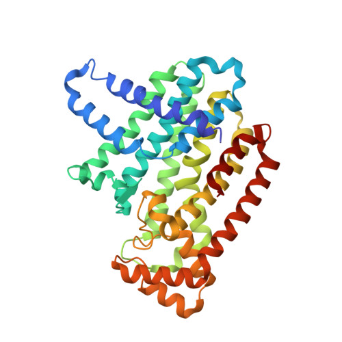
    Bisphosphonates are potent inhibitors of farnesyl pyrophosphate synthase (FPPS) and are highly efficacious in the treatment of bone diseases such as osteoporosis, Paget's disease and tumor-induced osteolysis. In addition, the potential for direct antitumor effects has been postulated on the basis of in vitro and in vivo studies and has recently been demonstrated clinically in early breast cancer patients treated with the potent bisphosphonate zoledronic acid. However, the high affinity of bisphosphonates for bone mineral seems suboptimal for the direct treatment of soft-tissue tumors. Here we report the discovery of the first potent non-bisphosphonate FPPS inhibitors. These new inhibitors bind to a previously unknown allosteric site on FPPS, which was identified by fragment-based approaches using NMR and X-ray crystallography. This allosteric and druggable pocket allows the development of a new generation of FPPS inhibitors that are optimized for direct antitumor effects in soft tissue.


  • Organizational Affiliation
    • Center for Proteomic Chemistry and Global Discovery Chemistry, Novartis Institutes for Biomedical Research, Basel, Switzerland. wolfgang.jahnke@novartis.com

Macromolecule Content 

  • Total Structure Weight: 40.92 kDa 
  • Atom Count: 2,958 
  • Modeled Residue Count: 343 
  • Deposited Residue Count: 350 
  • Unique protein chains: 1

Macromolecules

Find similar proteins by:|  3D Structure
Entity ID: 1
MoleculeChains  Sequence LengthOrganismDetailsImage
FARNESYL PYROPHOSPHATE SYNTHASEA [auth F]350Homo sapiensMutation(s): 0 
Gene Names: FDPSFPSKIAA1293
EC: 2.5.1.1 (PDB Primary Data), 2.5.1.10 (PDB Primary Data)
UniProt & NIH Common Fund Data Resources
Find proteins for P14324 (Homo sapiens)
Explore P14324 
Go to UniProtKB:  P14324
PHAROS:  P14324
GTEx:  ENSG00000160752 
Entity Groups
Sequence Clusters30% Identity50% Identity70% Identity90% Identity95% Identity100% Identity
UniProt GroupP14324
Sequence Annotations
Expand
Reference Sequence

Small Molecules

Ligands 4 Unique
IDChains Name / Formula / InChI Key2D Diagram3D Interactions
3N5

Query on 3N5


Download:Ideal Coordinates CCD File
B [auth F](2R)-1-[(benzyloxy)carbonyl]-2,3-dihydro-1H-indole-2-carboxylic acid
C17 H15 N O4
BSOYWTITVKXHLM-OAHLLOKOSA-N
ZOL

Query on ZOL


Download:Ideal Coordinates CCD File
C [auth F]ZOLEDRONIC ACID
C5 H10 N2 O7 P2
XRASPMIURGNCCH-UHFFFAOYSA-N
PO4

Query on PO4


Download:Ideal Coordinates CCD File
G [auth F]PHOSPHATE ION
O4 P
NBIIXXVUZAFLBC-UHFFFAOYSA-K
MG

Query on MG


Download:Ideal Coordinates CCD File
D [auth F],
E [auth F],
F
MAGNESIUM ION
Mg
JLVVSXFLKOJNIY-UHFFFAOYSA-N
Binding Affinity Annotations 
IDSourceBinding Affinity
ZOL BindingDB:  3N46 Ki: min: 0.07, max: 85.9 (nM) from 2 assay(s)
IC50: min: 0.24, max: 2000 (nM) from 18 assay(s)
3N5 BindingDB:  3N46 IC50: 5.00e+4 (nM) from 1 assay(s)

Experimental Data & Validation

Experimental Data

  • Method: X-RAY DIFFRACTION
  • Resolution: 2.35 Å
  • R-Value Free:  0.266 (Depositor), 0.266 (DCC) 
  • R-Value Work:  0.204 (Depositor), 0.204 (DCC) 
  • R-Value Observed: 0.210 (Depositor) 
Space Group: P 41 21 2
Unit Cell:
Length ( Å )Angle ( ˚ )
a = 110.853α = 90
b = 110.853β = 90
c = 70.861γ = 90
Software Package:
Software NamePurpose
XSCALEdata scaling
CNSrefinement
PDB_EXTRACTdata extraction
XDSdata scaling
XDSdata reduction
APRVphasing
CNXrefinement

Structure Validation

View Full Validation Report


Ligand Structure Quality Assessment 


Entry History 

Deposition Data

Revision History  (Full details and data files)

  • Version 1.0: 2010-08-18
    Type: Initial release
  • Version 1.1: 2011-07-13
    Changes: Version format compliance
  • Version 1.2: 2024-02-21
    Changes: Data collection, Database references, Derived calculations