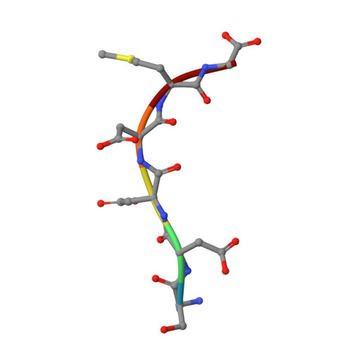
Crystal Structure of the complex formed between type I ribosome inactivating protein and hexapeptide Ser-Asp-Asp-Asp-Met-Gly at 1.7 A resolution
Kushwaha, G.S., Vikram, G., Sinha, M., Kaur, P., Sharma, S., Singh, T.P.To be published.
Experimental Data Snapshot
Starting Model: experimental
View more details
Entity ID: 1 | |||||
|---|---|---|---|---|---|
| Molecule | Chains | Sequence Length | Organism | Details | Image |
| Ribosome inactivating protein | 246 | Momordica balsamina | Mutation(s): 0 EC: 3.2.2.22 | 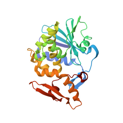 | |
UniProt | |||||
Entity Groups | |||||
| Sequence Clusters | 30% Identity50% Identity70% Identity90% Identity95% Identity100% Identity | ||||
| UniProt Group | D9J2T9 | ||||
Glycosylation | |||||
| Glycosylation Sites: 1 | |||||
Sequence AnnotationsExpand | |||||
Reference Sequence | |||||
| Ligands 1 Unique | |||||
|---|---|---|---|---|---|
| ID | Chains | Name / Formula / InChI Key | 2D Diagram | 3D Interactions | |
| GUN Download:Ideal Coordinates CCD File | D [auth A] | GUANINE C5 H5 N5 O UYTPUPDQBNUYGX-UHFFFAOYSA-N |  | ||
| Length ( Å ) | Angle ( ˚ ) |
|---|---|
| a = 130.241 | α = 90 |
| b = 130.241 | β = 90 |
| c = 39.974 | γ = 120 |
| Software Name | Purpose |
|---|---|
| HKL-2000 | data collection |
| AMoRE | phasing |
| REFMAC | refinement |
| DENZO | data reduction |
| SCALEPACK | data scaling |