3N33 | pdb_00003n33

Crystal structure of the N-terminal beta-aminopeptidase BapA in complex with pefabloc SC (AEBSF)


Experimental Data Snapshot

  • Method: X-RAY DIFFRACTION
  • Resolution: 1.80 Å
  • R-Value Free: 
    0.191 (Depositor), 0.185 (DCC) 
  • R-Value Work: 
    0.160 (Depositor), 0.155 (DCC) 
  • R-Value Observed: 
    0.160 (Depositor) 

Starting Model: experimental
View more details

wwPDB Validation 3D Report Full Report

Validation slider image for 3N33

Ligand Structure Quality Assessment 


This is version 1.3 of the entry. See complete history

Literature

Autoproteolytic and catalytic mechanisms for the beta-aminopeptidase BapA--a member of the Ntn hydrolase family.

Merz, T.Heck, T.Geueke, B.Mittl, P.R.Briand, C.Seebach, D.Kohler, H.P.Grutter, M.G.

(2012) Structure 20: 1850-1860

  • DOI: https://doi.org/10.1016/j.str.2012.07.017
  • Primary Citation Related Structures: 
    3N2W, 3N33, 3N5I

  • PubMed Abstract: 

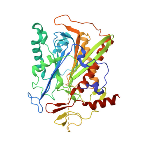
    The β-aminopeptidase BapA from Sphingosinicella xenopeptidilytica belongs to the N-terminal nucleophile (Ntn) hydrolases of the DmpA-like family and has the unprecedented property of cleaving N-terminal β-amino acid residues from peptides. We determined the crystal structures of the native (αβ)₄ heterooctamer and of the 153 kDa precursor homotetramer at a resolution of 1.45 and 1.8 Å, respectively. These structures together with mutational analyses strongly support mechanisms for autoproteolysis and catalysis that involve residues Ser250, Ser288, and Glu290. The autoproteolytic mechanism is different from the one so far described for Ntn hydrolases. The structures together with functional data also provide insight into the discriminating features of the active site cleft that determine substrate specificity.


  • Organizational Affiliation
    • Biochemistry Institute, University of Zürich, Winterthurerstrasse 190, 8057 Zürich, Switzerland.

Macromolecule Content 

  • Total Structure Weight: 158.55 kDa 
  • Atom Count: 12,211 
  • Modeled Residue Count: 1,468 
  • Deposited Residue Count: 1,492 
  • Unique protein chains: 1

Macromolecules

Find similar proteins by:|  3D Structure
Entity ID: 1
MoleculeChains  Sequence LengthOrganismDetailsImage
Beta-peptidyl aminopeptidase
A, B, C, D
373Sphingosinicella xenopeptidilyticaMutation(s): 0 
Gene Names: bapA
EC: 3.4.11.25
UniProt
Find proteins for Q52VH2 (Sphingosinicella xenopeptidilytica)
Explore Q52VH2 
Go to UniProtKB:  Q52VH2
Entity Groups
Sequence Clusters30% Identity50% Identity70% Identity90% Identity95% Identity100% Identity
UniProt GroupQ52VH2
Sequence Annotations
Expand
Reference Sequence

Small Molecules

Ligands 4 Unique
IDChains Name / Formula / InChI Key2D Diagram3D Interactions
EPE

Query on EPE


Download:Ideal Coordinates CCD File
F [auth A],
O [auth B]
4-(2-HYDROXYETHYL)-1-PIPERAZINE ETHANESULFONIC ACID
C8 H18 N2 O4 S
JKMHFZQWWAIEOD-UHFFFAOYSA-N
AES

Query on AES


Download:Ideal Coordinates CCD File
DA [auth D]
EA [auth D]
G [auth A]
H [auth A]
P [auth B]
DA [auth D],
EA [auth D],
G [auth A],
H [auth A],
P [auth B],
Q [auth B],
V [auth C],
W [auth C]
4-(2-AMINOETHYL)BENZENESULFONYL FLUORIDE
C8 H10 F N O2 S
MGSKVZWGBWPBTF-UHFFFAOYSA-N
SO4

Query on SO4


Download:Ideal Coordinates CCD File
AA [auth C]
BA [auth C]
FA [auth D]
GA [auth D]
HA [auth D]
AA [auth C],
BA [auth C],
FA [auth D],
GA [auth D],
HA [auth D],
I [auth A],
J [auth A],
K [auth A],
L [auth A],
M [auth A],
R [auth B],
S [auth B],
T [auth B],
X [auth C],
Y [auth C],
Z [auth C]
SULFATE ION
O4 S
QAOWNCQODCNURD-UHFFFAOYSA-L
GOL

Query on GOL


Download:Ideal Coordinates CCD File
CA [auth D],
E [auth A],
N [auth B],
U [auth C]
GLYCEROL
C3 H8 O3
PEDCQBHIVMGVHV-UHFFFAOYSA-N

Experimental Data & Validation

Experimental Data

  • Method: X-RAY DIFFRACTION
  • Resolution: 1.80 Å
  • R-Value Free:  0.191 (Depositor), 0.185 (DCC) 
  • R-Value Work:  0.160 (Depositor), 0.155 (DCC) 
  • R-Value Observed: 0.160 (Depositor) 
Space Group: P 1 21 1
Unit Cell:
Length ( Å )Angle ( ˚ )
a = 87.4α = 90
b = 96.85β = 108.38
c = 101.3γ = 90
Software Package:
Software NamePurpose
XDSdata scaling
PHASERphasing
PHENIXrefinement
XDSdata reduction
XSCALEdata scaling

Structure Validation

View Full Validation Report


Ligand Structure Quality Assessment 


Entry History 

Deposition Data

Revision History  (Full details and data files)

  • Version 1.0: 2011-06-08
    Type: Initial release
  • Version 1.1: 2011-07-13
    Changes: Version format compliance
  • Version 1.2: 2017-02-08
    Changes: Database references
  • Version 1.3: 2023-09-06
    Changes: Data collection, Database references, Derived calculations, Refinement description