3MX6 | pdb_00003mx6

Crystal structure of methionine aminopeptidase from Rickettsia prowazekii bound to methionine


Experimental Data Snapshot

  • Method: X-RAY DIFFRACTION
  • Resolution: 1.70 Å
  • R-Value Free: 
    0.199 (Depositor), 0.202 (DCC) 
  • R-Value Work: 
    0.160 (Depositor) 

Starting Model: experimental
View more details

wwPDB Validation 3D Report Full Report

Validation slider image for 3MX6

This is version 2.0 of the entry. See complete history

Literature

Rickettsia prowazekii methionine aminopeptidase as a promising target for the development of antibacterial agents.

Helgren, T.R.Chen, C.Wangtrakuldee, P.Edwards, T.E.Staker, B.L.Abendroth, J.Sankaran, B.Housley, N.A.Myler, P.J.Audia, J.P.Horn, J.R.Hagen, T.J.

(2017) Bioorg Med Chem 25: 813-824

  • DOI: https://doi.org/10.1016/j.bmc.2016.11.013
  • Primary Citation Related Structures: 
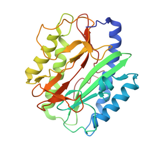
    3MR1, 3MX6

  • PubMed Abstract: 

    Methionine aminopeptidase (MetAP) is a class of ubiquitous enzymes essential for the survival of numerous bacterial species. These enzymes are responsible for the cleavage of N-terminal formyl-methionine initiators from nascent proteins to initiate post-translational modifications that are often essential to proper protein function. Thus, inhibition of MetAP activity has been implicated as a novel antibacterial target. We tested this idea in the present study by targeting the MetAP enzyme in the obligate intracellular pathogen Rickettsia prowazekii. We first identified potent RpMetAP inhibitory species by employing an in vitro enzymatic activity assay. The molecular docking program AutoDock was then utilized to compare published crystal structures of inhibited MetAP species to docked poses of RpMetAP. Based on these in silico and in vitro screens, a subset of 17 compounds was tested for inhibition of R. prowazekii growth in a pulmonary vascular endothelial cell (EC) culture infection model system. All compounds were tested over concentration ranges that were determined to be non-toxic to the ECs and 8 of the 17 compounds displayed substantial inhibition of R. prowazekii growth. These data highlight the therapeutic potential for inhibiting RpMetAP as a novel antimicrobial strategy and set the stage for future studies in pre-clinical animal models of infection.


  • Organizational Affiliation
    • Department of Chemistry and Biochemistry, Northern Illinois University, 1425 W. Lincoln Hwy, DeKalb, IL 60115, USA.

Macromolecule Content 

  • Total Structure Weight: 58.76 kDa 
  • Atom Count: 4,860 
  • Modeled Residue Count: 519 
  • Deposited Residue Count: 524 
  • Unique protein chains: 1

Macromolecules

Find similar proteins by:|  3D Structure
Entity ID: 1
MoleculeChains  Sequence LengthOrganismDetailsImage
Methionine aminopeptidase
A, B
262Rickettsia prowazekiiMutation(s): 0 
Gene Names: mapRP824
EC: 3.4.11.18
UniProt
Find proteins for Q9ZCD3 (Rickettsia prowazekii (strain Madrid E))
Explore Q9ZCD3 
Go to UniProtKB:  Q9ZCD3
Entity Groups
Sequence Clusters30% Identity50% Identity70% Identity90% Identity95% Identity100% Identity
UniProt GroupQ9ZCD3
Sequence Annotations
Expand
Reference Sequence

Small Molecules

Ligands 3 Unique
IDChains Name / Formula / InChI Key2D Diagram3D Interactions
MET

Query on MET


Download:Ideal Coordinates CCD File
D [auth A],
H [auth B]
METHIONINE
C5 H11 N O2 S
FFEARJCKVFRZRR-BYPYZUCNSA-N
FE

Query on FE


Download:Ideal Coordinates CCD File
E [auth A],
F [auth A],
I [auth B],
J [auth B]
FE (III) ION
Fe
VTLYFUHAOXGGBS-UHFFFAOYSA-N
NA

Query on NA


Download:Ideal Coordinates CCD File
C [auth A],
G [auth B]
SODIUM ION
Na
FKNQFGJONOIPTF-UHFFFAOYSA-N

Experimental Data & Validation

Experimental Data

  • Method: X-RAY DIFFRACTION
  • Resolution: 1.70 Å
  • R-Value Free:  0.199 (Depositor), 0.202 (DCC) 
  • R-Value Work:  0.160 (Depositor) 
Space Group: P 1 21 1
Unit Cell:
Length ( Å )Angle ( ˚ )
a = 50.37α = 90
b = 67.55β = 97.429
c = 80.89γ = 90
Software Package:
Software NamePurpose
REFMACrefinement
XSCALEdata scaling
PHASERphasing
PDB_EXTRACTdata extraction
XDSdata reduction

Structure Validation

View Full Validation Report


Entry History 

& Funding Information

Deposition Data


Funding OrganizationLocationGrant Number
National Institutes of Health/National Institute Of Allergy and Infectious Diseases (NIH/NIAID)United States--

Revision History  (Full details and data files)

  • Version 1.0: 2010-05-26
    Type: Initial release
  • Version 1.1: 2011-07-13
    Changes: Version format compliance
  • Version 1.2: 2017-02-08
    Changes: Database references
  • Version 1.3: 2017-02-15
    Changes: Database references
  • Version 1.4: 2023-09-06
    Changes: Data collection, Database references, Derived calculations, Refinement description
  • Version 2.0: 2026-03-11
    Type: Coordinate replacement
    Reason: Ligand identity
    Changes: Advisory, Atomic model, Author supporting evidence, Data collection, Database references, Derived calculations, Non-polymer description, Other, Refinement description, Source and taxonomy, Structure summary