3MX4 | pdb_00003mx4

DNA binding and cleavage by the GIY-YIG endonuclease R.Eco29KI inactive variant E142Q


Experimental Data Snapshot

  • Method: X-RAY DIFFRACTION
  • Resolution: 2.50 Å
  • R-Value Free: 
    0.274 (Depositor), 0.275 (DCC) 
  • R-Value Work: 
    0.211 (Depositor), 0.219 (DCC) 
  • R-Value Observed: 
    0.214 (Depositor) 

Starting Model: other
View more details

wwPDB Validation 3D Report Full Report

Validation slider image for 3MX4

This is version 1.3 of the entry. See complete history

Literature

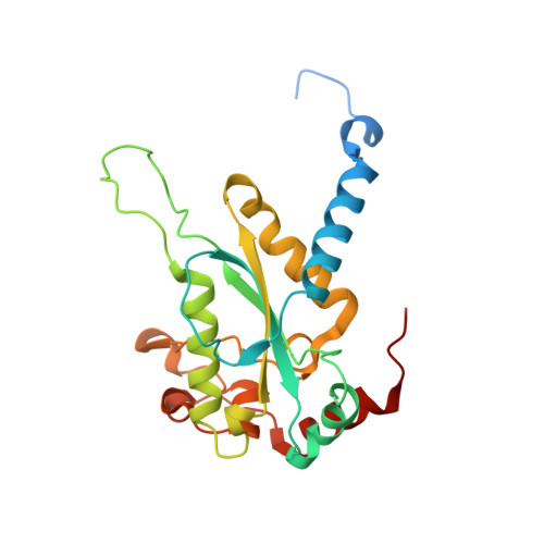
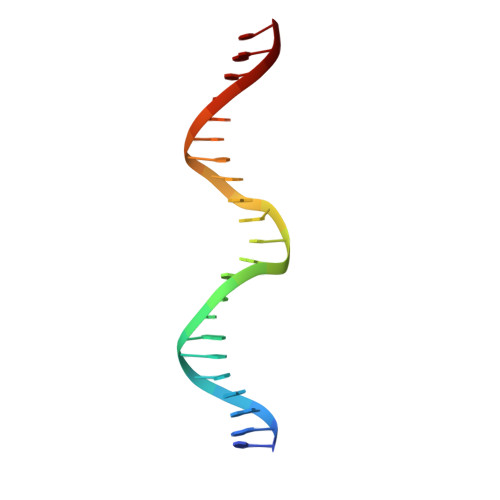
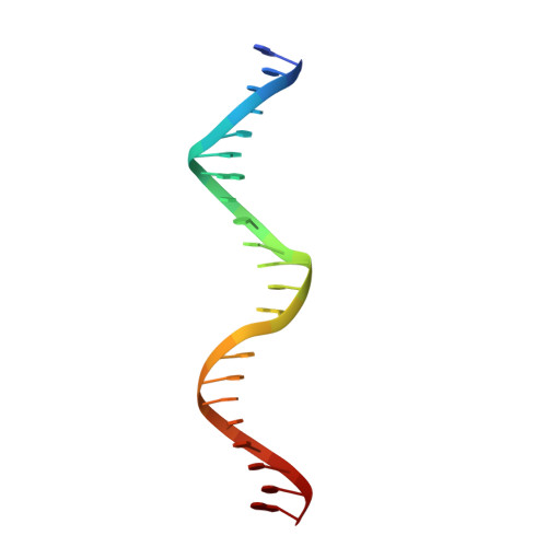
Folding, DNA Recognition, and Function of GIY-YIG Endonucleases: Crystal Structures of R.Eco29kI.

Mak, A.N.Lambert, A.R.Stoddard, B.L.

(2010) Structure 18: 1321-1331

  • DOI: https://doi.org/10.1016/j.str.2010.07.006
  • Primary Citation Related Structures: 
    3MX1, 3MX4, 3NIC

  • PubMed Abstract: 

    The GIY-YIG endonuclease family comprises hundreds of diverse proteins and a multitude of functions; none have been visualized bound to DNA. The structure of the GIY-YIG restriction endonuclease R.Eco29kI has been solved both alone and bound to its target site. The protein displays a domain-swapped homodimeric structure with several extended surface loops encircling the DNA. Only three side chains from each protein subunit contact DNA bases, two directly and one via a bridging solvent molecule. Both tyrosine residues within the GIY-YIG motif are positioned in the catalytic center near a putative nucleophilic water; the remainder of the active site resembles the HNH endonuclease family. The structure illustrates how the GIY-YIG scaffold has been adapted for the highly specific recognition of a DNA restriction site, in contrast to nonspecific DNA cleavage by GIY-YIG domains in homing endonucleases or structure-specific cleavage by DNA repair enzymes such as UvrC.


  • Organizational Affiliation
    • Division of Basic Sciences, Fred Hutchinson Cancer Research Center, Seattle, WA 98109, USA.

Macromolecule Content 

  • Total Structure Weight: 268.53 kDa 
  • Atom Count: 17,406 
  • Modeled Residue Count: 1,856 
  • Deposited Residue Count: 2,056 
  • Unique protein chains: 1
  • Unique nucleic acid chains: 2

Macromolecules


Find similar proteins by:|  3D Structure
Entity ID: 1
MoleculeChains  Sequence LengthOrganismDetailsImage
Eco29kIR
A, B, C, D, E
A, B, C, D, E, F, G, H
235Escherichia coliMutation(s): 2 
Gene Names: eco29kIR
EC: 3.1.21.4
UniProt
Find proteins for Q46944 (Escherichia coli)
Explore Q46944 
Go to UniProtKB:  Q46944
Entity Groups
Sequence Clusters30% Identity50% Identity70% Identity90% Identity95% Identity100% Identity
UniProt GroupQ46944
Sequence Annotations
Expand
Reference Sequence
Find similar nucleic acids by:  Sequence
Entity ID: 2
MoleculeChains LengthOrganismImage
DNA (5'-D(P*CP*GP*GP*GP*AP*GP*GP*CP*CP*CP*GP*CP*GP*GP*GP*CP*CP*GP*CP*CP*GP*C)-3')
I, K, M, O
22N/A
Sequence Annotations
Expand
Reference Sequence
Find similar nucleic acids by:  Sequence
Entity ID: 3
MoleculeChains LengthOrganismImage
DNA (5'-D(P*GP*CP*GP*GP*CP*GP*GP*CP*CP*CP*GP*CP*GP*GP*GP*CP*CP*TP*CP*CP*CP*G)-3')
J, L, N, P
22N/A
Sequence Annotations
Expand
Reference Sequence

Experimental Data & Validation

Experimental Data

  • Method: X-RAY DIFFRACTION
  • Resolution: 2.50 Å
  • R-Value Free:  0.274 (Depositor), 0.275 (DCC) 
  • R-Value Work:  0.211 (Depositor), 0.219 (DCC) 
  • R-Value Observed: 0.214 (Depositor) 
Space Group: P 1 21 1
Unit Cell:
Length ( Å )Angle ( ˚ )
a = 99.855α = 90
b = 101.463β = 110.19
c = 144.403γ = 90
Software Package:
Software NamePurpose
CrystalCleardata collection
PHASERphasing
REFMACrefinement
HKL-2000data reduction
SCALEPACKdata scaling

Structure Validation

View Full Validation Report


Entry History 

Deposition Data

Revision History  (Full details and data files)

  • Version 1.0: 2010-09-08
    Type: Initial release
  • Version 1.1: 2011-07-13
    Changes: Version format compliance
  • Version 1.2: 2024-02-21
    Changes: Data collection, Database references
  • Version 1.3: 2024-04-03
    Changes: Refinement description